
Subscribe to be the first to know about Best Deals and Exclusive Offers!
Interesting Cases in Echocardiography
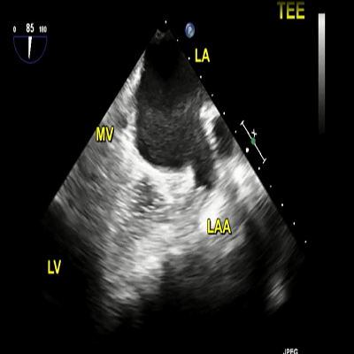
by Navin C NandaRight upper and lower pulmonary veins. Cropping the 3D dataset from the top shows infiltrated tissue on each side of RUP and RLP. LA, MV, AV, AO, LAA, SVC, IVC, and RVOT are also seen. Subsequent cropping from the side shows tumor tissue (arrows) in the IAS and between the RA and the confluent RPV. The pericardial effusion (hollow arrow) and pacemaker lead (arrowhead) are also seen.


© 2019 Jaypee Brothers Medical Publishers (P) LTD. | All Rights Reserved

 Refer to Friend
 Recommend To Librarian
