Manual for Dental Hygienist Maj Gen PN Awasthi
INDEX
Page numbers followed by b refer to box, f refer to figure, fc refer to flow chart, and t refer to table.
A
Abrasion 153, 154
Acellular cementum 87, 87f
Acetylated carbilols 121
Acidic sources, extrinsic 154
Acidogenic theory 176
Acids 125, 131, 176, 177
Acquired immune deficiency syndrome 165, 170
Acquired immunity 144
Acting agents, local 113
Actinobacillus actinomycetemcomitans 163, 220
Actinomyces 224
naeslundii 223
viscosus 223
Adequate nutrients within caloric needs 276
Adrenaline 113, 115
Agents 106, 108
for desensitization 135, 136
Aggressive periodontitis 212, 220, 221f
Air-powder polishing 245
Airway 298
Alcohol 121, 122, 124
Alcoholic beverages 277
Aldehydes 121
Alkalis 125, 131
Allergic reactions 212
Alveolar bone 47, 67, 87
proper 68
structure of 87f
Alveolar crest group 45
Alveolar mucosa 39
Alveolar nerve, inferior 28
Alveolar process 20, 26, 67
of mandible 68, 69f
of maxilla 68, 69f
Alveolar wall 6f
Alveoli 5
Amelogenesis imperfecta 150, 150f
American Academy of Periodontology 211
Aminoglycosides 109
Amoxicillin 111
Amphetamine 115
Ampicillin 111
Ampule 297
Amyl nitrate 116
Analgesic 119
effect 122
Anarthria 102
Anemia 156
Anesthesia
general 105
local 105, 107
surface 107
Anesthetic
effect 122
local 108
mouthwash 131
procedure 105
Angle's classification of malocclusion 66f
Angulation 243
Anodontia 149
Antibacterial function 94
Antibiotic 98
avoid use of 111
prophylaxis 111, 264
selection 110
usage 110
Anticalculus dentifrices 132
Anticaries dentifrice 132
Anticonvulsant effect 122
Antigen 139
Antigingivitis dentifrices 132
Antihemophilic globulin 113
deficiency of 157
Antiplaque dentifrices 132
Antiseptic 129, 188
Aphasia 102
Appliances, removable 266
Aramine 116
Areolar tissue 8, 9f
Arestin 133, 134
Aromatic food consumption 232
Arsenic trioxide 125, 126
Arthritis 233
Articular capsule 71
Articular disc 70
Ascorbic acid 278
Ascoxal 131
Aspirin 120
Astringents 131
Attrition 153, 153f
sign of 153
symptoms of 153
Augmentin 111
Autoimmunity 147
Automatic warning signal 196, 199
Axon 17
Azithromycin 111
B
Bacteria 110, 177
invisible to naked eye 127f
mechanical removal of 94
reservoir of 233
visible 127f
Bacterial antagonism 95
Bacterial endocarditis, subacute 233
Bacterial factors 214
Bacteroides
forsythus 163
melaninogenicus 224
species 163
Balanced occlusion 65
Bandage, four-tailed 286f
Barbiturate 121
poisoning, acute 122
Bass technique 252
Beam, attenuation of 198
Bell stage 82
advanced 82
Benzodiazepines 121, 122
Beta-lactum ring 109
Biohazard communication 190
Biomedical waste 189
management of 189, 189f
steps in 189
Bisecting angle technique 202, 202f
Bitewing 197
dental X-rays 197f
films 200
Black hairy tongue 170, 171f
Bladder 8f
Blood 13, 97, 114, 190
cells, types of 14f
coagulation 95
composition 97
dyscrasias 157
performs 98
pressure, rise in 117
smear 14f
supply 29, 72
interruption of 139
of palate 49, 50f
of teeth 48
tests, normal values of 99b
vessels 43
severed 112
Blood-borne pigmentations 228
Bocasin 132
Body 26
fracture of 289
mass index 281
Bone 13, 13f, 68
bundle 68
grafting, after 292
tissue 13
lacunae in 13f
types 68
wax 113
Borrelia vincentii 214
Breathing 298
Bristle angulation in
bass technique 252f
charters technique 253f
Bristle brush 245
Broad spectrum 109
Bronchial asthma 115
Brushing
chewing surfaces 249f
gums 266
manual versus automatic 248
Buccinator nerve 55
Bud stage 82
C
Calcium 114, 279
hydroxide 299
pyrophosphate 130, 254
requirements in pregnancy 282
sulfate 302
Calculus/deposits, removal of 266
Camphor 118, 124
Cancellous bone 67
Candida albicans 163
infection 156
Candida species 212
Candidiasis 165, 170, 171f
Canine 30, 32
Cap stage 82
Capnocytophaga 163
Carbohydrate 274, 277, 279
requirement 291
role of 277
sources 274
Carbolized resin 299
Carcinoma 173
Cardiopulmonary resuscitation 296, 298f
Cardiovascular system 122
Caries, smooth surface 178, 178f
Carious lesions, filling of 260
Carotid artery
branches of external 48, 49f
external 48
Caseous necrosis 143
Caustics 124, 125
Cautery, use of 113
Cavitron ultrasonic tips 239f
Cavity proper of mouth 21
CCD/CMOS sensor 205
Cefadroxil 111
Celecoxib 120
Cell 43, 68, 87, 88, 145
arrangement of 5f
blebbing 143
mediated immunity 145
membrane, cause leakage from 109
shape of 5f
structure of 3f
types of 41f
Cellular cementum 87, 87f
Cellular changes 143
Cellulose, oxidized 113
Cementoenamel junction 39, 85, 85f
Cementum 85, 87
Central incisor 30, 39f
Central nervous system 121
Centric occlusion 66
Centric relation 66
Cephalometry 199
of face 199f
Cervical caries 179, 179f
Cervical lymphadenopathy 216
Charters technique 252
Chediak-Higashi syndrome 221
Cheek 19
with mirror, retracting 241f
Chemical agents 139
Chemical materials 301
Chemical vapor 186
Chisel scaler 238, 238f
Chloral hydrate 123
Chloramphenicol 109
Chlorhexidine 132
chip, periochip 133, 134
Christmas disease 157
Chromic acid 125
Cilia 5
Ciprofloxacin 111
Circumpulpal dentin 86
Clarithromycin 111
Cleansing agent 129
Clindamycin 111
Clove oil 124
Coagulative necrosis 142
Coal tar derivatives 131
Cobalamin 278
Cold
chemicals 186
compress 112
sores 165
Coloring agents 130
Columnar epithelium, simple 5, 7f
Commensal flora 162
Communicability 215
Complete blood count 99b
Compressor naris 63
Condylar fracture 288
Condylar processes 28
Congenital syphilis 159
Connective tissue 7, 41f, 42
dense
irregular 10, 11f
regular 10, 11f
network of 9f
types of 8
Copper 279
Coronary heart disease 234
Coronoid process 28
fracture of 289
Corrugator supercilii 64
Cortical plate 67
Corynebacterium 163
Counterirritant 117
agents 117
type of 117
Coxsackievirus 164
Cranial nerves 48, 53, 54f
Craniomandibular joint 70
Cranium
forms bony protection for brain 29
muscle of 63
Creosote 124
Cribriform plate 68
Cuboidal epithelium, simple 5, 6f
Cyanocobalamin 159, 160, 218
Cyclo-propane 107
Cyst, after resection of 292
Cytotoxic hazard symbol 189f
D
Dead track 87
Dean's fluorosis
index 152t
stages of 151, 152f
Debilitating disease 214
Deciduous dentition 32
Defective fillings, correction of 260
Defense cells 88
Degeneration 142
Deglutition 100
stages 100
Dehydration 232
Demonstration 251
Dendrite 17
Dens in dente 149
Dental
amalgams 299
attrition 153
calculus 95, 225
classification 225
caries 96, 176, 232, 280
etiology 176
prevention of 179
chair after syncope 296f
Council of India 306
constitution of 306
functions of 306
curriculum 307
epithelium
inner 79
outer 80
ethics 305
films 200
uses 200
floss 256
unwaxed 256
waxed 256
follicle cells 77
health
and children 268
education 267
surveys 266
healthcare 181
histology 75
hygienist 261, 264, 295, 305
course 271
role of 269
working protocol of 261
hypersensitivity 135
junctions 86
lamina 78, 80
fate of 79
function of 78
management 175
materials 299
office 136, 296fc
antibiotic in 111
papilla 79, 83
personnel, protection of 166
pharmacology 103
physiology 75
plaque 96, 176, 223
induced gingival diseases 211
practice 181
antibiotic in 109
classification, antibiotic in 109
pulp 88
radiography 195
equipment, digital 205f
routine 198
radiology 193
research 307
sac 79
stains 227
tape 245
tissues
hard 84
soft 84
X-ray tube 196f
Dentalone 124
Dentifrices 129, 254
composition 254
special purpose 255
types 254
Dentin 85, 87
chemical composition 85
irregular 87
peritubular 86
primary 87
regular 87
sclerotic 87
secondary 87
structure of 86f
Dentinoenamel junction 85
Dentinogenesis imperfecta 150
type I 150f
Dentinopredentin junction 86
Dentist Act, 1948 and Dental Council of India 306
Dentistry
approach, preventive 306
history of 304
integration of preventive 307
preventive 265
public health aspect of 265
Dentoalveolar fracture 286, 288
Denture
artificial 232
base materials 300
care of 266
Deodorants 130
Deoxyribonucleic acid 279
Depressor anguli oris muscle 62
Depressor labii inferioris muscle 62
Dermis 15
Desensitizing agents 135
Desquamative gingivitis 217f
chronic 217
Detergent 129
Dettolin 132
Diabetes 159
mellitus 234
Diaminopyrimidines 109
Diclofenac 120
Diet 177, 276f
and dental caries 277
Dietary guidelines 276
Dietary reference intakes 275
Dietary sources 274
Digestive function 94
Digital radiography 204
Dilaceration 149, 149f
Disclosing agents 127, 128f
Disclosing solution 127
Disease Control and Prevention, centers for 185
Disinfect 184
Disinfectant 188
Disinfection 183
background of 183
Distal contacts 36
DNA function, interfere with 109
Domestic pressure cookers 187
Domestic water supplies, fluoridation of 267
Down's syndrome 221
Doxycycline 111
atridox 134
gel, atridox 133
Drug 119
allergy 110
delivery systems
advantages of local 134
disadvantages of local 134
indications of local 134
local 133
dependence 122
interaction 122
regimen 167
Dry heat 186
sterilizer 186f
Dysarthria 102
E
Eccrine gland 15
Elastic cartilage 11, 12f
Elastic impression materials 302
Elyzol 134
Enamel 84, 85
cuticle 77, 85
primary 85
secondary 85
formation completed 32
formed at birth 32
hypoplasia 150, 150f
rods 84
spindles 85
structure of 84f
tufts 85
Encouraged food groups 277
Endocrine 217
Endosteum 88
Endotoxins 146
Energy requirement 291
Entamoeba gingivalis 163
Environmental control 307
Enzymes 59, 146
Ephedrine 115
Epicranius muscle 63
Epidermis 15
Epithelium 3, 40
classification of 5f
Epsilon-aminocaproic acid 114
Epstein-Barr virus 163
Equipment 108
Ergot 116
Erosion 153, 154, 155f
etiology 154
Eruption 32, 150
delayed 150
sequence 30
time of 31
Erythema multiforme 166, 212
Erythrocytes 97
Erythromycin 109, 111
Esophageal stage 101
Esophagus 8f
Ether 106
Ethinamate 123
Ethyl chloride 106
Excretion 95
Exercises during pregnancy 283
Exotoxins 146
Exposing film 201
Extended shank curettes 236
Extraoral
films 201
fulcrums reinforcement 243
signs 215
symptoms 215
Extrinsic stains 227
Eyelid, muscle of 63
Eyes 122
F
Face, muscle of 36
Facial expression, muscle of 62
Facial muscles 62f, 64t
functional groups of 63t
Facial nerve 48, 53, 57
course of 57f
Facial paralysis 63
Fat 274, 277, 279
functions 274
necrosis 143
non-calorie roles of 274
requirements 292
Faulty brushing 247
Federation Dentaire Internationale 31
Feeding 293
Felypressin 116
Fiber 88
alveogingival 37
anterior 74
arrangement of principal 45
circular 38, 43
collagen 9, 43
dentogingival 37, 43
dentoperiosteal 37, 43
intercircular 38
intergingival 38
interpapillary 38, 45
tetracycline 133
transseptal 38, 43
Fibrin 113
Fibrinogen 113
Fibrinoid necrosis 143
Fibroblasts 43, 88
Fibrocartilage 11, 12f
Fibrous food, use of 266
File scaler 238, 238f
Filling materials 299
Film packets, placement of 198
Finger rest 242
on shank, pad of middle 242f
side of middle 242f
First-aid treatment 285
emergency 296f
Fissure caries 178
Fixation appliances, maintenance of 291
Flavoring agent 130
Floor of mouth 21, 22f
Flossing, technique of 256
Fluid 186, 190
exudates 140
Fluoride 130, 266, 279
ions 266
Fluorosis of teeth 151
etiology 151
histopathology 151
stages 151
treatment 152
Fluothane 106
Focal hyperkeratosis 170
Focal infection, mechanism of 232
Folate 278
requirements in pregnancy 282
Folic acid 160, 218, 278
Food
and nutrition 273
around teeth, retention of 231
classification of 273
impaction, correction of 260
safety 277
Food-borne illness 283
Fracture, site of 288f
Fungi 110
Fusion 149
Fusobacterium nucleatum 214, 224
Fusobacterium species 163
G
Galea aponeurotica 63
Gamma globulin 95
Ganglion 56
Gangrenous necrosis 143
Gas 186
in plasma 98
Gas-oxygen-ether technique 106
Gastrointestinal
diseases 234
tract 122
Gelfoam 113
Gemination 149
Genetic
disorders 212
factors 111
Genioglossus muscle 23
Germicide 188
Giant cell granuloma 219f
Gingiva 37
attached 38, 39
free 39
marginal 38
nerve supply to 52
parts of 40
types of 38, 38f
Gingival color 44
Gingival connective tissue 43
Gingival consistency 44
Gingival contour 44
Gingival crevicular fluid 43
Gingival diseases 211, 213, 258
acute 213
chronic 217
classification 209
management 209
Gingival fibers 43f
Gingival groove 39
Gingival lesions, non-plaque-induced 211
Gingival massage 225
Gingival overgrowth
classification 218
enlargement 218
Gingival position 44
Gingival sulcus 40
Gingival texture 44
Gingival tumors 218
Gingivitis 218, 232
causes of 266
plaque-induced 213f
prevention toothpastes, plaque and 254
with dental plaque 211
Gingivostomatitis, primary herpetic 164
Gland
classification of 89
mixed 24
structures 15
Glass bead 186
Glenoid fossa 74
Glycerine 130
Goblet cells 5
Gracey curettes, modifications of 236
Gram-negative
cocci 162
rods 163
Gram-positive
cocci 162
filaments 163
Granulation tissue 140
Gums 20
Gutta percha 299
H
Habitually open mouth 266
Halitosis 231
from nasopharynx 232
Halothane 106
Handling and disposing sharps 190
Haversian system 68
Hazards 189
Health
education 268
effects on general 232
general 276f
Healthy gingiva 44f
tissue 39f
Heart, acceleration of 117
Hematologic diseases 156
Hematologic problems 217
Hematological disorders 212
Hemophilia 157
Hemorrhage 285
Hemostatic agents 112, 113
Hepatic function 110
Hepatitis 167
A 167, 168
B 167
prevalence of 168
virus 163
C 167
virus 169
D 167
virus 169
delta virus 169
E 167, 169
treating patients with 169
Herpes encephalitis 164
Herpes labialis 164f
Herpes simplex virus 164, 165
Herpetic gingivostomatitis 164f
acute 216, 216f
diagnosis, acute 216
treatment, acute 216
Hirschfeld's canals 67
Hoe scalers 238
Host defense, impaired 111
Human
anatomic waste 190
anatomy 1
body, tissues in 3
dentition 30
eruption of 32t
immunodeficiency virus 165, 170, 181
papilloma viruses 164, 165
tissues 190
trachea 7f
Humectants 130
Humoral immunity 145
Hunter-Schreger bands 85
Hyaline cartilage 11, 12f
Hydrocolloids alginate, irreversible 302
Hyoglossus muscle 23
Hypersensitivity 147
Hypnotics classification 121
Hypodermis 15
Hypoplasia 148
Hypotension 115
I
Iatrogenic factors 178
Ibuprofen 120
Idiopathic resorption 36
Immature bone 68
Immune response 144
abnormalities of 147
role of 147
Immunity 144
active
artificial 144
natural 144
passive
artificial 144
natural 144
Immunoglobulins 146
Immunologic problems 217
Implants, plastic instruments for 238
Impression materials 300
classification 300
Incisor
first 32
labial surface of 35f
lateral 30
peg-shaped lateral 148f
second 32
Indomethacin 120
Infection
after 292
control 184
protocol for 181
methods of controlling 290
transmitted 183
Infectious disease hazard communication 190
Infectious hepatitis 168
Infiltration, local 107
Inflammation 139
acute 139
causes of
acute 139
chronic 140
chronic 140
fate of
acute 140
chronic 140
of gums 211
stages of acute 139
Inflammatory enlargement 218f, 219
chronic 219
treatment of 219
Infrabony pocket 222, 222f
Infrahyoid muscles of mastication 60f
Inhibin 95
Inhibit cell wall synthesis 109
Inhibit DNA gyrase 109
Inhibit protein synthesis 109
Injectable emergency drugs
primary 297t
secondary 297t
Injection 297
Inorganic
ions 123
substance 84, 98
Instrument
condition of 241
grasp 242
handling 242
maintenance of 188
Insulin 264
Integument 15
Intercellular
ground substance 43
matrix 87
substance 88
Interdental
brushes 257
cleaning aids 255, 266
gingiva 38, 40
pointed 39f
Interlobular dentin 86
Intermaxillary fixation 285
Inter-rod substance 85
Interseptal injections 107
Intertubular dentin 86
Intra-articular disc 72
Intraoral
finger rests 243
variations in 243
radiography 197
films, types of 197
Intraosseous injections 107
Intrinsic acidic sources 154
Intrinsic stains 228
Iodine 118
Iron 279
deficiency anemia 282
in pregnancy 282
treatment 282
Irrigation 290
Isoprenaline 115
Isoproterenol 115
J
Jaundiced eyes 168f
Jaw
anatomy of 25
ectomesenchyme of 82
immobilization, after 293
Junctional epithelium 42
K
Kaposi sarcoma 165
Karyolysis 143
Karyorrhexis 143
Keratinized gingiva 39
Ketamine 106
Ketoprofen 120
Kidney 122
tubules 6f
Korff's fiber 88
L
Labial frenulum
lower 19f
upper 19f
Labiolingual 78f
Lactic acid 125
Lactobacillus acidophilus 177
Lamellated Haversian system 67
Langer curettes 238
Laughing muscle 62
Leukemia 156, 221
Leukemic gingival enlargements 219
Leukocyte 13, 97
adhesion deficiency syndrome 221
characteristics of 98b
Leukoderma 170
Leukoplakia 170, 171f
Leukotoxin 94
Levator anguli oris muscle 62
Levator labii superioris 62
alaeque nasi muscle 62
Lichen planus 170, 171f, 212
Lignocaine hydrochloride 108
Linea alba 19, 20f
Lingual frenulum 22f
Lingual nerve 57
Linoleic acid 274
Linseed-mustard 117
Lip 19, 19f
everted
lower 19f
upper 19f
lower 62
with index finger, retracting 241f
Liquefactive necrosis 143
Liquor soda ethylate 125
Listeria 163
monocytogenes 283
Listerine 132
Ludwig's angina 234
Lung disease 232
Lupus erythematosus 212
Lymph 13
nodes of head and neck 14f
Lymphocyte 43, 145
Lysozyme 95
M
Macrodontia 148
Macrolides 109
Magnesium 279
aluminum silicate 130, 254
Malignant lesion, after resection of 292
Malocclusion 65
class II 66
classification of 66
Malpractice 305
common grounds for 305
criteria for 305
defence against 305
Mandible 26
age changes in 29, 29f
adult 29f
child 29f
old 29f
fracture of 286
inner surface of 27f
outer surface of 27f
Mandibular
condyle 70
division 55
fossa 71
fracture 287
classification 287
distribution of 288f
etiology of 288
site of 288, 288f
type of 287
teeth 65
in oral cavity, positioning of 35f
nerve supply of 51, 51f, 51t
Mantle dentin 86
Manual processing 204, 204f
Masseter muscle 58, 60, 60f
deep part 60
functions 60
superficial part 60
Mast cells 43
Mastication 100
muscle of 59, 59f
Materials, variety of 299
Mature bone 68
Maxilla 25
fracture of 286
lateral aspect of 25f
medial aspect of 26f
Maxillary
artery 28, 49f
branches 49f
division 53
internal 56
fractures 286
classification 286, 287f
sinus 69
teeth 65, 250f
in oral cavity, positioning of 35f
nerve supply of 50, 50f, 50t
Maxillofacial
injury 285, 290, 293
transportation of 291
surgery 284, 292
McCall's festoon 230
Mean corpuscular
hemoglobin 99
volume 99
Medical emergencies, management of 295, 296fc
Medicated dentifrices 132
Meissner's corpuscle 15
Meloxicam 120
Menstrual breath 232
Mentalis muscle 62
Menthol 118, 124
Mesial contacts 36
Mesiobuccal 50
Mesiodistal section 47f
Metabolic diseases 232
Metabolism, interfere with intermediary 109
Metal ions 132
Metallic 300
salts 126
stain 228
Metaraminol 116
Methedrine 116
Methohexital 107
Methoxyflurane 107
Methyloxathiazinone dioxide 254
Metronidazole gel 111, 133, 134
Micrococcus 162
Microdontia 148
Microorganisms 139, 163
group of 162
in mineralization, role of 227
Microvilli 5
Minerals 275
deficiency, symptoms of 278t
Mini-bladed curettes 237
Mini-langer curettes 238
Minocycline 134
microspheres 133
Molar 33, 51
first 30, 32
occlusal surface of 35f
second 30, 32
upper 20f
Morphine 119
Mouth
breathing 232
muscle of 62
Mouthwashes 129, 131, 254
Movements, types of 73
mRNA code, cause misreading of 109
Mucocutaneous lesions 212
Mucogingival
deformities 213
junction 39
Mucosa 19, 190
nerve supply to 52
Mucosal burns 170
Mucous
glands 24
membrane 167t
Muscle structures 15
Mutinase 95
Mycoplasma species 224
Mylohyoid 73
nerve 57
N
Nasal decongestion 115
Nasal-inferior nasal 56
Nasalis muscle 62
Nasmyth's membrane 77
Nasogastric tube feeding 293
Nasotracheal intubation 105
Neck, muscle of 63
Necrosis
etiology of 142
types of 142
Necrotizing ulcerative gingivitis, acute 131, 213, 214f
Neisseria 162
gonorrhoeae 211
Neodymium-doped yttrium aluminium garnet 136
Neonatal line 86
Neoplasm 173
Neoplastic enlargement 218
Nerve 43
block 107
endings, free 15
Nervous
control 102
structures 15
tissue 15
Neuron, structure of typical 17f
Neutropenia 221
Niacin 218, 278
Nicotinamide 159
Nicotinic acid 160
Nimesulide 120
Nitric
acid 125
oxide 188
Nitrobenzenes 109
Nitroimidazoles 109
Nitrous oxide 106
Non-injectable emergency drugs
primary 297t
secondary 297t
Nonmetallic 300
stain 228
Nonoperating hand 241f
Nonselective CoX-2 inhibitors 120
Nonsharp disposable items 190
Nonsteroidal anti-inflammatory drugs 119, 120
Noradrenaline 115
Nose, muscle of 62
Nostrils 63
Nutrients 273, 279, 280
deficiency, symptoms of 278t
Nutrition 291
and health 275
and periodontal diseases 280
during pregnancy and lactation 281
feeding 291
Nutritional
assessment 292
deficiency 214, 217
needs during pregnancy 281
requirements 292
status 276f
O
Obtundents 124
types 124, 125
Occipitofrontalis muscle 63
Occlusal 198
films 201
forces, insufficient 231
radiograph 198f
trauma 213
Occlusion 65
functional 65
ideal 65
Octapressin 116
Ocular diseases 234
Odontoblasts 88
Odontogenic infection, distant spread of 234
Operation theater protocol 293
Opioids 119
newer 119
Opsonin 94
Oral
and maxillofacial surgery 284
cancer 173
classification 174
etiology 174
management of 175
staging of 175, 175t
carcinomas, staging of 175
cavity 18
bacteriology of 162
proper 18f
sagittal section of 18f
virology of 162
white lesions of 170, 170t, 172, 172fc
epithelium 41, 41f
feeding 293
fissure 63
foci of infection 233
significance of 233
health 276f
hygiene
aids 235, 246
methods of performing 253
status 265
immunology 144
acquired 144
innate 144
types 144
irrigation devices 225
malignancy 173
mucositis 156
prophylaxis 258, 259, 265
goals of 260
sepsis 232
signs 214
squamous cell carcinoma 172
stage 100
submucous fibrosis 170, 172
surgery 284
symptoms 215, 216
viral
infections, common 164
leukoplakia 165
Orbicularis oculi 63, 64
Orbicularis oris 63, 64
muscle 62
Organic substances 84, 98
Organisms, pathogenicity of 146
Orthodontic, preventive 260
Orthopantomographs 201
Osseous tissue 13, 13f
Ossification 29
Osteocytes 13
Oxidizing 131
Oxycel 113
P
Pacinian corpuscle 15
Pain 153, 179
sensations during oral prophylaxis, control of 264
Palatal surfaces, strokes for 250f
Palate
hard 21
and soft 21f
nerve supply of 49, 50f
soft 21
Palatine
middle 56
posterior 56
process 26
Palatine nerve
greater 49
lesser 50
Palatoglossus muscle 23
Palm and thumb grasp 242f
Palpebral fissure 63
Panoramic radiograph 199f
Pantothenic acid 159, 160
Papilloma 166
Papillon-Lefèvre syndrome 221
Paracetamol 120
Paraformaldehyde 125
plus zinc oxide 125
Paraldehyde 123
Paralleling technique 202
Paraperiosteal injections 107
Parasymphysis, fracture of 289
Parenteral nutrition 293
Parotid
fascia 63
glands 28
Patient and operator, protection of 196
Pemphigoid 212
Pemphigus vulgaris 212
Pen grasp
modified 242f
standard 242f
Penicillin 109
Pentazocine 119
Peptococcus 162
Peptostreptococcus 162
Percutaneous injuries 167t
Periapical
films 200
radiographs, techniques for 201
X-ray exposure 197f
Pericoronitis 216
management 217
Periocline 133, 134
Periodontal
anatomy 37
disease 147, 211, 220, 233f, 234, 258
classification 209
management 209
necrotizing 212
signs 220
symptoms 220
types of 220
untreated 211
instrument
classification of 235
parts of 235f
ligament 44, 46
fibers 45f
pocket 221
classification of 222f
probes 235
surgical kit 240f, 241
treatment 264
Periodontitis 212, 221, 232, 266
chronic 212, 220
generalized 220f
necrotizing 221f
with endodontic lesions 212
Periodontium 37
abscesses of 212
hard tissues of 37
structure of 37f
to occlusal forces 231
Periosteogingival fibers 38
Periosteum 88
Permanent filling materials 299
Permanent teeth 30, 31f
Pethidine 119
Pharyngeal stage 101
Phenol 124, 126
Phentolamine 116
Phenytoin-associated enlargement 218f
Phonation 100, 101
Phosphorus 279
sensor 205
Physical agents 139
Physical method 112
Piroxicam 120
Pit and fissure caries 178f
Plaque
accumulation, causes of 225
formation of 224
marginal 223
Plaque control 224
instructions 225
mechanical 224
methods of 224
Plaque removal
frequency of 225
technique for 224
Plasma 98, 114
cells 43
Plaster of Paris 300
Plaster, uses of 301
Plastic probes 238f
Platelets 13
Platysma 64
muscle 63
Polishing 261
Polishing cloth 258
Polishing methods 245
Polishing teeth, procedure for 246
Polycythemia 157
Polypeptides 109
Poor oral hygiene 177
Porous loads 186
Porphyromonas gingivalis 163
Potassium 277
bromide 123
hydroxide 126
salt 254
of 6-methyloxathiazinone dioxide 130
thiocyanate 116
Prazosin 116
Predentin 86
Pregnancy 177
outcome 234
Pregnant woman
increased nutritional risk in 281
weight gain chart in 281t
Premature eruption 150
Premolars 51
Pressure 112
Prilocaine 108
Priscol 116
Procerus muscle 62
Processing film 203
developing 203
fixing 203
washing 203
Professional negligence 305
Proliferative verrucous leukoplakia 172
Prophylactic fillings 260
Prophylactic regimen, standard 111
Prophylactic restoration 265
Prophylaxis, post-exposure 166, 167
Propofol 107
Protective foods 280
Protein 274, 279
deficiency 218
functions 274
nutrition status, assessment of 274
requirement 291
Proteolysis-chelation theory 176
Proteolytic theory 176
Protozoa 163
Protrusion 74
Pseudohemophilia 157
Pseudostratified columnar epithelium 6, 7f
Psychic flow 93
Psychosomatic factors 214
Pterygoid 56
function
lateral 61
medial 61
muscle
external 61
internal 61
lateral 61, 61f, 28
medial 61, 61f
Pterygomandibular raphe 27
Public health, concept of 267
Pustulants 117
Pyknosis 143
Pyridoxine 159, 160, 218, 278
Q
Quinolones 109
R
Radiation 187
protection supervisor 196
warning sign 196
Radiological protection authority 198
Ramus 26, 28
fracture of 289
Reaction, type of 145
Recommended dietary allowance 276
Recurrent aphthous ulceration 166
Red blood cell 97, 97f, 98, 99, 279
Red cell distribution width 99
Refined carbohydrate consumption, regulation of 179
Removing debris 253
Renal diseases 234
Renal function 110
Resorption, external 36
Resting flow 93
Reticular tissue 10, 10f
Retromolar trigone 20
Retrusion 74
Retzius, incremental lines of 85
Reversible hydrocolloids 301
Riboflavin 159, 160, 218, 278
Ribonucleic acid 279
Rigid materials 301
Risorius muscle 62
Rod sheath 84
Roll technique 252
Root formation completed 32
Root planing 262
Root sensitivity 135
Root, development of 80f
Rubber base impression materials 302
Rubber cup 244
S
Saliva 92, 95, 177
amount of 92
buffering power of 95
chemical composition 92
functions 92
in disease 95
in health 94
putrification of 232
Salivary enzymes, role of 96
Salivary flow, factors affecting 93
Salivary gland 24, 89, 91
classification of 24, 89
duct system, structure of 91f
major 24, 90f
types of 90t
Salivation, mechanism of 93
Sarcoma 173
Scaling 261
Sclerosing agents 114
Scurvy 161
symptoms of 161f
Secretory cells 91
types of 93
Selective CoX-2 inhibitors 120
Selenomonas species 163
Semicircular fibers 38
Semicylindrical structure 70
Sensitivity 153
Serous glands 24
Serum 167
hepatitis 168
Shallow rapid respiration 117
Sharpening, principles of 244
Sharpey's fibers 46, 88
Silver nitrate 125
Skeletal muscles 62
Skin
diseases 234
exposure, non-intact 167t
structure of 16f
Smokers' breath 232
Snake venom 114
Socket wall 88
Sodium 126, 277, 302
carboxymethyl cellulose 130, 254
coconut monoglyceride sulfonate 130, 254
lauryl sulfate 130, 254
phosphate 302
Soft tissue, adjacent 112, 250f
Soiled dressings 190
Soiled wastes 190
Sonic scalers 239
Sorbitol 130
Speech, disorders of 102
Sphenopalatine 56
Spinal cord 122
Spirochaetes 224
Squamous cell carcinoma 165
Squamous epithelium
simple 5, 6f
stratified 6, 8f
Stannous fluoride 130, 254
Staphylococcus 162
salivarius 162
Steam sterilization 185f
Steel ball 186
Stellate reticulum 79
Sterilization
background of 183
indicators of 187
methods of 185
unacceptable methods of 187
uses of 183
Sterilize 184
Stillman's cleft 230
Stippling, absence of 230
Stomach mucosa 7f
Stratum intermedium 79
Streptococcal viridans 234
Streptococci 162
Streptococcus
milleri 162
mitior 162
mitis 223
mutans 162, 177, 223, 277
salivarius 162, 177, 223
sanguinis 177
sanguis 162, 223
Streptomycin 109
Strokes 243
Styloglossus muscle 23
Stylohyoid muscle 57
Subgingival
calculus 226, 226f, 262
environment 233
plaque 223
Sublingual fold 19
Sublingual gland 19
Sublingual papilla 19, 22f
Submucosal injection 107
Sulcular and junctional epithelium 42f
Sulcular epithelium 42
Sulfanilamide 109
Suprabony pocket 221, 222f
Supragingival calculus 226, 226f
Supragingival plaque 223
Surgery, art of 284
Sweetener 254
Sweetening agent 130
Symphysis, fracture of 289
Syncope
signs of 295
symptoms of 295
Synovial cavities 71
Syphilis 157, 158
primary stage 157
secondary stage 158
symptoms of stages 158f
tertiary stage 158
Systemic acting agents 113
Systemic disease 156
manifestation of 212, 221
T
Tannic acid 113
Taurodontism 149
Teeth 20, 32, 50, 100, 178, 190, 233f, 249f, 258f
anatomy, loss of 153
anomalies of 148
anterior 51, 150f
buccal aspect of 43f
bud 80
section of 78f
damage to 250f
development of 77
stages 80
extraction, after 292
functions of 31
germ 77
growth 77
stages in 80t, 81f
impacted 150
lower posterior 249f
nerve supply of 48
numbering system 31
types of 34f
universal 33f
of lower jaws 25
of upper jaws 25
pathological migration of 230, 230f
polishing of 244, 245
position, maintenance of 35
primary 30, 30f
primordial of 80
resorption of 36
secondary 30
supernumerary 149
surfaces, natural 245
type of physiological 153
upper
anterior 249f
posterior 249f
whitening toothpastes 254
Temporalis muscle 60, 60f
function 61
insertion of 61f
Temporary filling materials 299
Temporomandibular joint 60, 70, 71, 73
anatomy of 71, 71f
movement of 73f
structure 70
Temporoparietal muscle 63
Tetracyclines 109
Thermoplastic materials 300
Thiamine 159, 218
Thickening agents 130
Thiopentone 106
sodium 106
Thrombin 113
Thrombocytes 98
Thrombocytopenic purpura 157
Thromboplastin 113
Thymol 124
Tincture aconite 118
Tinidazole 111
Tissue
adipose 10, 10f
classification of 3, 4f
types of 4f
Tobacco
cessation 269
stains 228f
Tongue 22, 90
coated 232
dorsum of 23f
geographic 170
muscle of 22, 23f, 36
scraper 258, 258f
ventral surface of 22f
Toothbrush
and toothbrushing 246
choice of 247
types of 247f
use of automatic 248f
Toothbrushing techniques 248
Tooth-colored filling materials 299
Toothpaste 130, 254
anti-caries/cavity protection 254
composition of 130t, 254t
formula 130
types of 254, 254t
Tourniquet 112
Trabeculae 91
Tramadol 119
Transgingival fibers 38
Transitional epithelium 7, 8f
Transseptal group 45
Trauma from occlusion 229
acute 229
chronic 229
primary 229
secondary 229
Traumatic crescent 230
Traumatic lesions 212
Treponema
denticola 163
macrodentium 163
oralis 163
pallidum 157, 211
Trichloroacetic acid 125
Trichloroethylene 107
Trichomonas 163
Trigeminal nerve 48, 53, 55f
branches of 56f
Tuberculosis 143, 159
Tumor
benign 173, 292
malignant 173
types of 173
Turpentine oil 118
U
Ultrasonic scalers 239
advantages 239
disadvantages 241
types 239
Urea derivatives 121
V
Varicella-zoster virus 163
Vascular response 139
Vascular supply 46
Vasoconstrictors 115
Vasodilators 115, 116
Veillonella species 162
Venous return from teeth and jaws 52
Verruca vulgaris 164
Vesicants 117
Vessels 28
Vestibular lamina 79
Vestibule
lower 19f
upper 19f
Villi 5
Virology 163
Viruses, classification of 163
Visibility 241
Vitamin 275
A 159, 218, 278, 292
deficiency of 159
and mineral in pregnancy 282
B 218, 292
complex 159
B12 278
B2 278
B3 278
B6 278
B9 278
C 114, 160, 218, 292
D 161, 278
deficiency 159, 218
symptoms of 278t
E 278
K 114, 278, 292
requirements 292
Volkmann's canals 67
W
Wafers 127
use of 128f
Waste
category 189, 190t
sharps 190
survey 189
type of 190
Water 279
and minors 130
balance 95
boilers 187
irrigation outfits 258
Waxes and resins, impression 301
Wearing occlusal splint 154f
Weight gain, rate of 281
White blood cells 98, 99, 279
emigration of 139
types of 98f
White sponge nevus 170
Wood stick 256, 257f
Wound healing 161
X
Xerostomia 166
Xylocaine, topical 264
Z
Zinc 279
chloride 124
oxide 299, 302
eugenol 299, 302
phosphate cements 299
Zygomatic process 26
Zygomaticus muscles
major 62
minor 62
×
Chapter Notes

Save Clear


1Human Anatomy
SECTION OUTLINE
  • → Tissues in Human Body
  • → Oral Cavity
  • → Anatomy of Jaws
  • → Human Dentition: Primary and Permanent Teeth
  • → Periodontal Anatomy: Clinical and Histological Characteristics
  • → Blood and Nerve Supply of Teeth and Associated Structures
  • → Cranial Nerves: Trigeminal and Facial Nerve
  • → Muscles of Mastication
  • → Muscles of Facial Expressions
  • → Occlusion and Malocclusion
  • → Alveolar Process
  • → Temporomandibular Joint2

Tissues in Human BodyCHAPTER 1

 
INTRODUCTION
Cells are the smallest units of life. Cell is defined as the structural and functional unit of the living body. Clusters of cells are formed according to the comparable structure and function, which in turn leads to the formation of tissues in complex organisms. These tissues act as a unit and aid in various functioning of the organs which are essential to maintain biological life.
The current chapter sheds light on histology, the study of various tissues found in the human body along with its composition and function.
The structure of the cell contains (Fig. 1.1):
  • Cell membrane
  • Nucleus
  • Cytoplasm containing different organelles.
 
CLASSIFICATION OF TISSUES
The human body is made up of four basic types of tissues depending on their composition and function; namely epithelium, connective, muscular, and nervous tissues (Fig. 1.2).
The different function of each tissue is as follows:
  • Epithelium: Lines and covers surfaces
  • Connective tissue: Protects, supports, and binds together
  • Muscular tissue: Produces movement
  • Nervous tissue: Receive stimuli and conduct impulses.
zoom view
Fig. 1.1: Structure of the cell
 
EPITHELIUM
It forms the covering of surface of the body and aids in protection, adsorption, excretion, secretion, filtration, and sensory reception. However, it is important to consider the following points, which are as follows:
  • Arrangement of epithelium is done to make one surface free and one surface attached, known as apical surface and basal surface, respectively
  • Cells in epithelium are arranged either side by side or atop each other to form sheets of cells which in turn are held together by specialized junctions
  • Basement membrane is formed by the attachment of epithelium to a layer of connective tissue at the basal surface. This adhesive layer is basically composed of secretions from the epithelial cells and the connective tissue cells
  • Epithelium does not have its own blood supply4
zoom view
Fig. 1.2: Classification of four basic types of tissues
  • However, cells of epithelium can be regenerated, if properly nourished.
 
Classification
Depending upon the basis of shape of the cells and the arrangement of the cells within the tissue, the epithelium is divided (Figs 1.3A and B). Generally, the arrangement of the cells is stated first, then the shape which is followed by epithelium to complete the naming, e.g. simple squamous epithelium.
According to the arrangement of the cells:
  • Simple: Cells are found in a single layer attached to the basement membrane
  • Stratified: Cells are found in two or more layers arranged atop each other
  • Pseudostratified: A single layer of cells which appears to be multiple layers due to difference in height and location of the nuclei in the cells5
zoom view
Fig. 1.3A: Classification of epithelium on the basis of arrangement of cells
zoom view
Fig. 1.3B: Classification of epithelium on the basis of shape of cells
  • Transitional: Cells are round in shape and can slide across one another to allow stretching.
According to the shape:
  • Squamous: Flat, thin, scale-like cells
  • Cuboidal: Cells have a basic cube shape in which the cell's height and width are about equal
  • Columnar: Tall, rectangular or column-shaped cells which are basically taller in comparison to their width.
 
Distinguished Features
  • Cilia (singular: cilium, Latin: eyelash): These are hair-like appendages which attaches to the apical surface of cells to produce movement
  • Goblet cells: Specialized cells secrete mucus for lubrication and protect the surface of an organ
  • Villi (singular: villus, Latin: shaggy hair): Some organ exhibits finger-like projections that arise from the epithelial layer to increase the surface area for faster and more efficient adsorption
  • Microvilli: Small projections that arise from the cell's surface also help in enhancing surface area for absorption. Based on their bushy appearance, they are sometimes called as brush border of an organ.
 
Simple Squamous Epithelium
Simple squamous epithelium, single layer of flattened cells lines small air sacs of lungs called alveoli (Fig. 1.4A). One surface of epithelium faces an open space to the inside of the air sac, known as lumen and the cell surface facing it is the apical surface.
 
Simple Cuboidal Epithelium
Simple cuboidal epithelium lines kidney tubules and appears as dispersed thick-walled circular structures, when seen in the histological cross section (Fig. 1.4B).
 
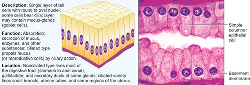
Simple Columnar Epithelium
Simple columnar epithelium covers the lining of the intestine.6
zoom view
Fig. 1.4A: Simple squamous epithelium in alveolar wall
zoom view
Fig. 1.4B: Simple cuboidal epithelium in kidney tubules
The lining extends into the lumen and is usually seen as finger-like projections called villi. These villi aids in enhancing the absorption of the intestine by increasing the surface area. On cross section, along with large rectangular cells, clear-looking goblet cells are also found which secretes mucus for protection of the intestinal lining (Fig. 1.4C).
 
Pseudostratified Columnar Epithelium
This type of epithelium is typified by irregularly shaped cells which mark the lining of trachea. The nuclei of the cells are seen scattered in different spots within the cells, which provide the tissue its false-layered appearance. In addition, it is covered with numerous tiny hair-like structures called cilia, which trap debris and dispense mucus secreted by the goblet cells across the surface of the tissue (Fig. 1.4D).
 
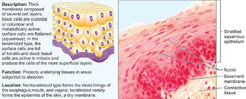
Stratified Squamous Epithelium
Epidermis, the outer layer of skin constitutes multiple layers of flattened cells. However, these cells get flattened and subsequently die as the cells are pushed higher. The death of the cell is attributed to the depletion of nutrients from the sources present in the connective tissue. These, in turn, form the outer dead layers of the epidermis (Fig. 1.4E).7
zoom view
Fig. 1.4C: Simple columnar epithelium in stomach mucosa
zoom view
Fig. 1.4D: Pseudostratified columnar epithelium in human trachea
 
Transitional Epithelium
This type of epithelium resembles a combination of cuboidal and columnar shaped cells. This is seen in the inner lining of the bladder in which tissue can stretch and distend without causing perforations in the wall lining (Fig. 1.4F).
 
CONNECTIVE TISSUE
It is the most extensively scattered tissue type present in the human body. It helps in protecting, supporting and binding together parts of the body.
The following section throws some light on the chief characteristics of connective tissue:
  • Though connective tissue is extremely vascularized, some tissues such as tendons, ligaments, and cartilages are less vascular in nature
  • It is composed of various types of specialized cells
  • It encompasses a huge amount of non-living material known as the matrix in the form of ground substance and fibers, which in turn are formed and secreted by the cells of the specific connective tissues.8
zoom view
Fig. 1.4E: Stratified squamous epithelium in esophagus
zoom view
Fig. 1.4F: Transitional epithelium in bladder
 
Classification
There are four types of connective tissues found in the human body (Fig. 1.5A):
  • Connective tissue proper
    • Loose connective tissue
      • Areolar
      • Adipose
      • Reticular.
    • Dense connective tissue
      • Dense regular
      • Dense irregular.
  • Supporting connective tissue
    • Cartilage
      • Elastic
      • Hyaline
      • Fibrocartilage.
    • Bone
  • Fluid connective tissue
    • Blood
    • Lymph.
 
Areolar Tissue
It is a loose type of connective tissue which constitutes intermixing of fibers and cells. It shows a very diffuse arrangement (Fig. 1.5B). The cells appearing as dark spots within the tissue are known as fibroblasts (fibro: fiber, blast: to create).9
zoom view
Fig. 1.5A: Network of connective tissues
zoom view
Fig. 1.5B: Areolar tissue
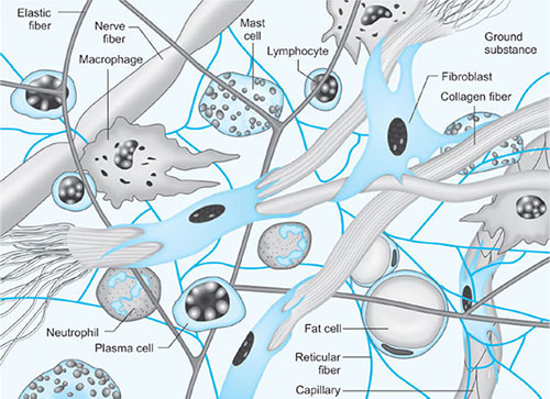
The prime function of these cells is to secrete the fibers. The larger fibers which appear pink in color and small black fibers are called collagen fibers and elastic fibers, respectively. Both these fibers and the rest of the substance collectively surround the cells, known as matrix.10
zoom view
Fig. 1.5C: Adipose tissue
zoom view
Fig. 1.5D: Reticular tissue
 
Adipose Tissue
These tissues present as large, irregular, bubble-like cells clustered together and scattered in a matrix alike areolar tissue (Fig. 1.5C). The cells are occupied with fat which acts as storage of energy, insulation, and protection.
 
Reticular Tissue
These fibers aid in supporting the cells of a soft internal skeleton like spleen, lymph nodes, and bone marrow (Fig. 1.5D).
 
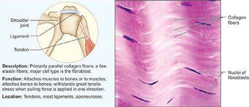
Dense Regular Connective Tissue
This tissue is surrounded by the fibers and plays an important role of producing fibers. In addition, it also forms tendons and ligaments that are required to connect muscles to bones and bones to other bones (Fig. 1.5E).
 
Dense Irregular Connective Tissue
Dermis, the middle layer of the skin constitutes this type of tissue. Though composition is homogenous as of dense regular tissue, its constituents are not arranged appropriately as compared to dense regular tissue (Fig. 1.5F).11
zoom view
Fig. 1.5E: Dense regular connective tissue
zoom view
Fig. 1.5F: Dense irregular connective tissue
 
Hyaline Cartilage
The lining below trachea is composed of rings of hyaline cartilage which are immersed in the organ in order to offer structure and support. The cartilage on cross section shows smooth, amorphous matrix containing collagen fibers with cells embedded within it. This matrix is produced by the cells known as chondrocytes which are contained within tiny chambers called lacunae or little lakes (Fig. 1.6).
 
Elastic Cartilage
The tissue that needs more stretching and maintenance of shape contains elastic fibers within the matrix of the tissue (Fig. 1.7). This type of tissue is present majorly in the structure of the external ear.
 
Fibrocartilage
Its arrangement is comparable to hyaline cartilage and forms the intervertebral discs in the spinal column, the pubic symphysis, and in the discs of the knee. It differs from hyaline cartilage in its rigidity, as it is less firm and exhibits more number of collagen fibers (Fig. 1.8).12
zoom view
Fig. 1.6: Hyaline cartilage
zoom view
Fig. 1.7: Elastic cartilage
zoom view
Fig. 1.8: Fibrocartilage
13
zoom view
Fig. 1.9: Bone or osseous tissue
zoom view
Fig. 1.10: Lacunae in bone tissue
The cells found are also known as chondrocytes which are found in the lacunae.
 
Bone (Osseous) Tissue
Bone tissue is organized in concentric ring structures called osteons in center of which lies a Haversian canal (Fig. 1.9A). This canal contains blood vessels and provides nutrition to the cells within the tissue.
Moreover, this canal is surrounded by rings of bone tissue called lamellae which contain chambers as lacunae (Fig. 1.10). These lacunae in turn possess tiny channels called as canaliculi which permit the interaction of the cells with the blood supply. The cells embedded in the tissue are known as osteocytes (osteo: bone, cyte: cell).
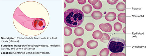
 
Blood
Blood is wrongly not considered as a tissue due to its liquid state, yet it fulfills all the requirements of being a connective tissue. It contains chiefly two types of cells; red blood cells (erythrocytes) and white blood cells (leukocytes).
In addition, tiny fragments of cells called platelets are also seen in this tissue (Fig. 1.11). Along with these cells, matrix also known as plasma is present which is made up of water, salts, gases, and proteins (nonliving materials).
 
Lymph
It is a thin opalescent fluid originating in organs and tissues of the body that circulates through the lymphatic vessels and is filtered by the lymph nodes. It fight infection, and in which are formed blood cells that is lymphocytes, monocytes, and plasma cells (Figs 1.12A and B).14
zoom view
Fig. 1.11: Blood smear showing various types of blood cells
zoom view
Figs 1.12A and B: Lymph nodes of head and neck (A) Anatomical locations; (B) Histology
15
Lymphatic system is a vast, complex network of capillaries, thin vessels, ducts, nodes and organs that help to protect and maintain the internal fluid environment of the entire body.
 
THE INTEGUMENT
The skin or integument is the main organ which protects the body externally. Besides protection, it plays a vital role in providing insulation, temperature regulation and tactility (sense of touch). It also aids in the formation of vitamin D, essential for proper functioning of the body.
 
Composition
The skin is composed of three distinctive layers namely; epidermis, dermis and hypodermis.
 
Epidermis
It is the outer layer which is formed of keratinized stratified squamous epithelium, which is arranged into five layers called strata (singular: stratum), which are shown in Figs 1.13A and B)
  • Stratum corneum, which is the outermost layer of flattened, dead cells
  • Stratum lucidum, thin translucent layer present only in thick areas of the skin
  • Stratum granulosum is named because of its richness of granules. The cells begin to die in the upper boundary of this layer
  • Stratum spinosum is one of the thicker layers of dermis with potential of fast division of cells
  • Stratum basale is the lowest layer of the skin which is attached to the dermis to form basement membrane. In this layer, cells continuously divide to create new cells.
 
Dermis
It is the middle layer and is formed from dense irregular connective tissue.
Dermal papillae, projections or ridges arise from the dermis and offer attachment points for the epidermis.
 
Hypodermis
It is the innermost or subcutaneous layer which is composed of adipose tissue.
 
Various Structures and Functions
The distinctive structures and functions of skin are as follows:
  • Gland structures
    • Eccrine gland: It secretes sweat which is the combination of water, salts, and urea and helps in cooling down the temperature of the body
    • Sebaceous gland: It secretes sebum or oil which aids in keeping the skin soft and pliable.
  • Nervous structures
    • Free nerve endings: These are positioned near dermal papillae and are related with pain sensation
    • Meissner's corpuscle: These touch receptors are positioned near dermal papillae and are associated with tactility
    • Pacinian corpuscle: These pressure receptors are situated deep within dermis at the boundary of the dermis and hypodermis.
  • Muscle structures
    • Arrector pili: These are the muscle that pulls up hair follicle causing goose flesh or goose bumps.
  • Appendages
    • Hair shaft
    • Hair follicle.
 
NERVOUS TISSUE
Neurons are the smallest functional units of the nervous system. Each neuron is composed of a cell body and two types of processes: axon and dendrites (Fig. 1.14). The function of the neurons is reflected in the number and types of processes arising from the cell body.16
zoom view
Figs 1.13A and B: Structure of skin
17
zoom view
Fig. 1.14: Structure of a typical neuron
 
Axon
It conducts impulses to other neurons or cells. Neurons have only one axon. The cell membranes of myelin cell predominantly contain fat which gives brain's white matter its fatty appearance.
 
Dendrite
It conducts impulses from synapse to the cell body. Depending on its function, a neuron may have multiple dendrites. Dendrites may undergo complex arborization to increase their surface area.