Pocket Tutor Surface Anatomy Richard Tunstall, S. Ali Mirjalili
INDEX
Note: Page numbers in bold or italic refer to tables or figures respectively.
A
Abdomen 199200
bony landmarks 200204
borders of 200, 201
dermatomes 213
duodenum 226, 227, 228
four-region model 222, 222
gallbladder 224, 225, 226
gastrooesophageal junction 225, 227
inguinal canal 210, 210213, 212
intestine 337338
intra-abdominal vasculature 213, 213216, 215
liver 224, 224, 225
liver and gallbladder 339
mesenteries and omenta 339
muscles, tendons and aponeuroses 207210, 208, 209
nine-region model 222223
oesophagus 225, 225
pancreas 226, 227, 228
reference planes 202, 203, 205
regional pain, localisation of 223, 223
regions 205, 205, 206
spleen 226, 227, 229
stomach 226, 228, 337
visceral contents of regions 222, 222223
Abdominal aorta 215216
anterior branches 215, 215, 216
lateral branches 215216
terminal branches 216
Abdominal aortic aneurysms (AAAs) 215
Abdominal wall, neurovasculature of 217, 217219, 218
arteries 217, 218219
lymphatic drainage 219
Abdominal wall, surgical access via 219221
incisions 219221, 220
trochar insertion 221
Abduction and adduction 11, 1213
finger 13
hip 12
shoulder 12
thumb 13
toe 13
wrist 13
Abductor digiti minimi 315, 316
Abductor hallucis 315, 316
Abductor pollicis longus 139
Acromioclavicular joint 124
Acromion 122
Adduction 11 see also Abduction and adduction
Adductor brevis 276, 277
Adductor canal 281283
Adductor hiatus 275277, 276
Adductor longus 274, 276
Adductor magnus 275277, 276
Allen test 146
Anal canal 245247, 247
Anal triangle 241242, 242, 242, 243
Anatomical movements 7
abduction and adduction 11, 1213
eye movements 21, 21
flexion and extension 7, 711, 11
inversion and eversion of foot 16, 1617
lateral flexion 20, 20
mandibular movements 17, 17
opposition and reposition of digits 19, 19
pronation and supination 15, 1516, 16
rotation 14, 1415
scapular movements 17, 18
Anatomical planes 4, 6, 6
Anatomical position 3, 4
Anatomical snuffbox 154156, 155, 156
Anconeus 139
Anconeus triangle 138, 139, 140
Ankle
bones of 308309, 314
dorsal 313
ligaments 309, 309, 310
retinacula of 310311, 311, 312
Anserine bursa 280, 281
Anterior ethmoidal artery 75
Anterior shoulder dislocation 124
Anterior superior iliac spine 204, 237, 237
Anterior thigh (Holden's) skin crease 264, 266
Anterior thoracotomy incisions 96
Anterior tibial artery 302, 304
Appendix 231
Arm
deep veins and lymph nodes of 135
muscles 130, 131
neurovasculature of 133
Arteries 331332
Axial plane 6, 6
Axilla, neurovasculature of 133
Axillary artery 132, 134
Axillary lines and folds 94, 96, 9698, 97
anterior axillary line 98
midaxillary line 98
posterior axillary line 98
safe zone/triangle 96, 97
Axillary lymph nodes 102104, 103
anterior (pectoral) nodes 103
apical nodes 104
central nodes 104
lateral (humeral) nodes 103
posterior (subscapular) nodes 103
Axillary vein 133, 135, 135136
B
Back 169 see also Vertebral column
Back, muscles of
deep and thoracolumbar fascia 191193, 192
deep intrinsic layer 193, 193, 196
intermediate extrinsic layer 191, 192
posterior view 192, 194, 196
superficial extrinsic layer 189191, 190
Baker cysts 297
Basilic vein 135, 136, 143, 147, 148, 149
Bell clapper deformity 258
Biceps brachii 130
Biceps femoris 274, 275
Biceps femoris tendon 290
Biceps tendon 143
Bicipital aponeurosis 143
Bladder 252, 253
Brachial artery 135, 141, 143, 144, 144, 145
Brachial plexus and branches 7071, 125, 130, 132, 132, 133
Brachiocephalic veins 116
Brachioradialis 141
Brain 343
lobes, cerebellum and dural venous sinuses 4041, 41, 42
sulci, fissures and gyri 3940, 40, 41
Breast 104105, 105
areola and nipple 104105, 105
axillary tail of 104
size, shape and position of 104
Buccinator 44, 45
Bulbourethral glands 255, 256
Bursa of gluteal region, thigh and knee 280, 280, 281
C
Caecum 230231
Calcaneal (Achilles) tendon rupture 293
Calcaneal tuberosity and processes 308
Calcaneonavicular (spring) ligament 309
Capitulum 136
Cardiac tamponade 100
Carotid bifurcation 76, 80
Carpal bones 150, 151, 152, 153, 153
Carpal tunnel 153, 159, 160, 165
Carpal tunnel syndrome 160
Carpometacarpal joint 154, 155
Cauda equina 185, 186, 188
Cauda equina compression 188
Cauda equina syndrome 294
Caudal epidural anaesthesia 173
Central sulcus of brain 39, 40
Cephalic vein 103, 104, 135, 136, 143, 147, 148, 149, 155
Cerebellum 42
Cervical lordosis 182
Cervical lymph node, deep 82, 83
Cervical spinal nerves 49, 50
Cervical spine, sagittal MRI 181
Chest wall deformities 87
Ciliary glands 58, 59
Circumflex humeral arteries 134
Circumflex iliac arteries 219
Clavicle 88, 89, 124
Clitoris 249, 249250, 250
Coccydynia 179
Coeliac plexus block 172
Colles’ fascia 244, 244
Colles’ fracture 149
Colon 230, 231, 231233
Common carotid artery 7576
Common fibular nerve 299, 301, 303, 304
Compartment syndrome 121, 263
Condylar line 39
Conjoint tendon 207
Conjunctival sac and fornix 58, 60
Coracoacromial ligament 126, 127
Coracoclavicular ligaments 126, 127
Coracoid process 88, 89, 122
Cornea 58, 60
Coronal plane 6, 6
Corpus cavernosum 254, 254255
Corpus spongiosum 254, 255
Costal cartilages 9192
Costal margin 92
Costoclavicular ligament 126, 127
Costodiaphragmatic recess 107
Costotransverse joints 178, 178
Cranial fossae 41, 4142
anterior cranial fossa 41
middle cranial fossa 42
posterior cranial fossa 42
Cranium 27
neurocranium 27, 27
reference lines of 39, 39, 40
viscerocranium 27, 27
Cubital fossa 141, 142, 143, 144
Cubital lymph nodes 143
Curvatures, vertebral column 179, 181, 181, 182
Cutaneous branches of cervical plexus 70, 73
Cutaneous innervation 22
dermatomes 22, 22, 23
lower limb 25
upper limb 24
Cutaneous nerves and arteries 102
C1 vertebra 34
D
Dacryocystocele 61
Deep fibular nerve 305, 316317
Deltoid 128, 129
Deltoid ligament 309
Deltoid tubercle 123
Deltopectoral lymph nodes 103, 104
Deltopectoral triangle 130
Depressor anguli oris 44
De Quervain's tenosynovitis 156
Dermatomes 22, 22, 23
abdominal 213
pelvic girdle and perineum 261
thoracic 100
Diaphragm 94, 200, 326, 327
Digastric muscle 67
Digital arteries and nerves 168
Digital extensor tendons 158
Digital pulp spaces 162, 163
Digital synovial sheaths 161
Dimple of Venus 204
Dinner fork deformity 149
Dorsal interossei 158
Dorsalis pedis artery 316, 319
Dorsal metatarsal arteries 318319
Dry eye 43
Dural venous sinuses 40, 41
E
Echocardiography 116
Elbow 136, 137
humerus 136
joint 137, 138
ligaments 139
radius 138
superior radioulnar joint 138139
ulna 137138
Emergency thoracotomy 09
Epiphora 43, 60
Epistaxis 46
Erector spinae 194, 193, 193, 195
Ethmoidal sinus 47, 47, 48, 48
Ethmoidal sinusitis 47
Eversion 16, 16
Extension 7, 11 see also Flexion and extension
Extensor carpi radialis longus 139
Extensor digitorum brevis 313
Extensor digitorum longus 299, 300
Extensor hallucis brevis 313
Extensor hallucis longus 299, 300
Extensor muscles 139, 140, 141
Extensor pollicis brevis 139
External carotid artery 76, 77
External jugular vein 76, 77
External oblique 207
External pudendal arteries 219
Eye
ciliary glands 59
conjunctival sac and fornix 58, 60
cornea and sclera 58, 60
eyelids 57, 58
lacrimal duct system 59, 59, 6061
lacrimal gland 59, 59
lens of eye 58, 60
movements 21, 21
ocular ultrasound 58, 60
tarsal glands 59
Eyeball trauma 60
F
Facet/zygapophyseal joints 178, 179
Facial nerve (CN VII) 52, 5355, 54, 56
Facial skeleton and mandible 343344, 343
Felon 162
Female external genitalia
bulbs of vestibule 250, 250251
clitoris 249, 249250, 250
greater vestibular glands 250, 251
labia majora 248, 248
labia minora 249, 249
mons pubis 248, 248
urethra 251
vaginal vestibule 249, 249251, 250
Female pelvis
bladder 252, 253
median sagittal MRI 253
uterus 251
vagina 251, 252
Female perineum
ischioanal fossae 245, 246
neurovasculature and pudendal canal 259
pelvic diameters 240, 241
pelvic floor muscles 245
perineal fascia and membrane 244
urogenital and anal triangles of 241, 242
Femoral artery 281, 283286
Femoral hernias 204, 281
Femoral neck fractures 272
Femoral nerve 284, 285, 286
Femoral sheath 280281
Femoral triangle 279, 282, 283
Femoral vein 281
Femur 269270, 271
greater trochanter 269270
lateral femoral condyles 270
medial femoral condyles 270
shaft 270
Fibula 289290
Fibular artery 302, 303
Fibularis brevis 301, 302
Fibularis longus 299300, 301
Fibularis tertius 299, 300
Fibula trochlea 308
Finger invagination test 211
Flexion and extension 7, 711, 11
ankle 10
digital 9
elbow 8
hip 9
knee 10
neck 10
shoulder 7
thumb 9
toe 10
vertebral column 11
wrist 8
Flexor carpi radialis 143, 160
Flexor carpi ulnaris 143, 160
Flexor digitorum brevis 315, 316
Flexor digitorum longus 296, 297
Flexor digitorum profundus 159, 160
Flexor digitorum superficialis 143, 159, 160
Flexor hallucis brevis and sesamoid bones 315
Flexor hallucis longus 296, 297, 314
Flexor muscles 139, 141, 142, 143
Flexor retinaculum 310
Fontanelles 38
Foot
bones of 307308, 308, 309, 310
dorsal 313, 314
plantar 313314, 315, 316, 317, 318
tarsal bones and joints of 308
tendons and retinacula of 311, 312
Forearm 121
muscles and tendons 139, 140, 141, 142, 143
neurovasculature of 144
superficial veins and cutaneous nerves of 148
Frontal bone 29, 3032
Frontal lobe 42
Frontal sinus 47, 47, 48, 48
G
Gallbladder 224, 225, 226
Ganglia, on extensor carpi radialis brevis tendon 156
Gastrocnemius 294, 295, 295
Gastrooesophageal junction 225, 227
Genitourinary system 339, 340
Glans of clitoris 250, 250
Gluteal crease 266, 267
Gluteal region injection 267269, 269, 270
dorsogluteal 268269
ventrogluteal 268
Gluteal triangle 268
Gluteus maximus 272273, 273
Gluteus medius 273, 273
Gluteus minimus 273, 273
Golfer's elbow 136, 141
Gracilis 276, 277
Greater supraclavicular fossa 70
Greater vestibular glands 250, 251
Guyon's canal 164, 165, 167
H
Haemorrhoids 247
Halitosis 64
Hallux valgus 308
Hamstring muscle group 274, 275
Hand
bones 149, 150, 151, 152, 153, 155
carpal bones 150, 151, 152, 153, 153
dorsal 158
dorsal venous network of 147, 147
metacarpals 150, 151, 154, 155
palmar 158, 160161, 165, 166
phalanges 154
right hand, arteriogram of 167
synovial sheaths and spaces of 161163, 162
Head and neck
anterior fontanelle 342
arteries 7376, 74
bones and bony landmarks 2938, 3032, 3537
ectopic neck viscera and neck lumps 8485, 86
head 27, 27
intracranial structures 39, 3942, 40, 41, 42
larynx and trachea 79, 80, 81
location of 343
lymphatics 8184, 82, 83, 84
muscles of head and face 4246, 43
neck 2728, 28
nerves and nerve point of neck 7073, 71, 72, 73
nerves of face 4955, 50, 5052, 54
nose and nasal cavity 46, 47
oral cavity and oral vestibule 6164
paranasal sinuses 47, 47, 48, 48
posterior fontanelle 342343
thyroid gland 81
triangles of neck 6570, 66, 67, 68, 69, 69
veins 7678, 78
vertebral levels 28, 29
viscera of face 5561, 5659
Head of talus 308
Heart 111116, 113, 114, 115, 330
aorta 112, 114, 115, 116, 118
borders 111112, 113, 114, 115
great vessels 112, 113, 114
valves and auscultation points 116, 117
veins 113, 114, 116
Hip joint 271, 272, 272
Holden's crease 208, 208
Hook of hamate 152, 153, 153
Houseman's vein see Cephalic vein
Humerus 123, 123124, 125, 136
greater tubercle 124
intertubercular sulcus 124
lesser tubercle 124
spiral/radial groove 124
surgical neck 124
Hydrocele 258
Hyphema 60
Hypoglossal nerve 73
Hypothenar eminence 159, 160
I
Iliac bone 202, 202, 204
Iliac crest 204, 236, 237, 237
highest point of 237, 237238
Iliac tubercle 204, 237, 237
Iliotibial tract 263264, 265
Iliotibial tract (band) syndrome 264
Ilium 237, 237238
Index–metacarpal–pisiform line 152, 153
Infant calvaria 38
Inferior alveolar nerve 51, 53
Inferior angle 123
Inferior belly of omohyoid 68
Inferior epigastric artery 218219
Inferior extensor retinaculum 310, 311
Inferior fibular retinaculum 311, 311
Inferior lumbar (Petit's) triangle 190, 191
Inferior ramus 238
Inferior vena cava 216
Infraclavicular fossa 130
Infraorbital nerve 49, 51, 52, 53
Infrapatellar bursa 280, 281
Infrapatellar nerves 305, 306
Infraspinatus 128, 129
Inguinal canal 210, 210213, 212
contents of 212, 212213
deep inguinal ring 211
hernia formation 211
midinguinal point 211
midpoint of inguinal ligament 211
superficial inguinal ring 211
Inguinal ligament 208
Intercostal muscles 93, 93
Intercostal nerves 102, 103
Intercostal neurovascular bundles 100102, 101
Intercostal spaces 88, 92
Intermediate cutaneous nerve 317
Internal jugular vein 76
Internal oblique 207
Internal pudendal artery 259, 259260
Internal thoracic artery and vein 102
Interspinous ligament 179, 180
Interspinous plane 202, 203
Interspinous spaces 170
Inversion 16, 16
Ischial bursa 280, 280
Ischial spines 237, 238
Ischial tuberosity 237, 238
Ischioanal fossae 235, 245, 246
Ischium 238
J
Jain's point 221
Jugular venous pulse 76
Jugulodigastric nodes 83
K
Kidneys 232, 233, 233234
Kiesselbach's plexus 46
Knee joint 291, 291293
injection/aspiration 279
lateral femoral condyle 291, 291
ligaments 292293, 293, 294
medial femoral condyle 291, 291
menisci 291, 292
popliteal fossa 297, 298, 299
tendons 292, 292, 293, 294
Kyphosis 181, 182
L
Labia majora 248, 248
Labia minora 249, 249
Lacrimal duct system 59, 59, 6061
Lacrimal puncta 60
Lacrimal sac 60
Laminae 178, 173
Laparoscopic ports 221
Large intestine 230, 231
Laryngeal inlet 79, 81
Larynx 79, 80
body of the hyoid 79, 80
cricoid cartilage 79
cricothyroid membrane 79
thyrohyoid membrane 79
thyroid cartilage laminae 79
Lateral (fibular) collateral ligament 292293
Lateral (radial) collateral ligament 139
Lateral cutaneous nerve of thigh 286, 287
Lateral epicondyle 136, 137
Lateral flexion 20
neck 20, 20
vertebral column 20, 20
Lateral meniscus 291, 292
Lateral rotator muscle group 273
Lateral (Sylvian) sulcus of brain 39, 40
Latissimus dorsi 128, 129
Lee-Huang's point 221
Lesser supraclavicular fossa 70
Levator ani 244245
Levator labii superioris 44
Ligamentum flavum 179, 180
Ligamentum nuchae 179, 180
Linea alba 208, 209, 209
Linea semilunaris 210
Lingual nerve 64
Little's area 46
Liver 224, 224, 225, 226
Long extensor muscle tendons 313, 314
Long thoracic nerve 102
Lordosis 181, 182
Lower limb 263
ankle and foot 307319
arteries and veins 281284, 283286
fascia and compartments 263, 263, 264266
femur 269270, 271
hip joint 271, 272, 272
iliotibial tract 263264, 265
knee, popliteal fossa and leg 289307
muscles, tendons and regions 272281
nerves 284287, 285, 286, 288
patella 270271
pelvic girdle 267269, 271
skin creases 265266, 267
Lumbar lordosis 182
Lumbar puncture 188, 188189
Lumbar spine
sagittal MRI 172
ultrasound 172
Lungs and pleura 105111, 106, 108, 109
borders of lung and pleura 106, 107, 108
horizontal fissure 110
lobes and fissures 106, 108, 109110, 111
needle decompression 108
oblique fissure 110
pleura and costodiaphragmatic recess 105107, 106
surface markings of 107, 108, 109
thoracostomy 108109
tracheobronchial tree 110111, 112, 113
Lymphadenopathy 81
Lymph nodes of head and neck 8184, 82, 83, 84, 85
M
Male external genitalia
bulbourethral glands 255, 256
corpus cavernosum 254, 254255
corpus spongiosum 254, 255
neurovasculature of dorsal penis 260
penis 253, 253256
scrotum 256
spermatic cord 258
swellings, testicle/scrotum 258
testicles 256258, 257, 258
urethra 255, 256
Mandible 31, 33
alveolar process 33
angle 33
body 33
coronoid process 33
mandibular notch 33
ramus 33
Mandibular movements 17
protrusion 17, 17
retrusion 17, 17
Mandibular nerve 33
Masseter 46
Mastoid line 39
Mastoid process 344
Maxilla 30, 3133
alveolar processes 33
body 32
frontal process 32
palatine processes 33
Maxillary nerve 33
Maxillary sinus 47, 47, 48, 48
McBurney's point 220, 221
Medial (tibial) collateral ligament 293
Medial (ulnar) collateral ligament 139
Medial cutaneous nerve 318
Medial epicondyle 136, 137
Medial meniscus 291, 292
Median cubital vein 148, 149
Median nerve 142, 143, 144, 146, 164, 165
Median vein of the forearm 148, 149
Mediastinum 98100, 99
anterior 100
location and borders 98, 99
middle 100
posterior 100
superior and inferior 9899, 99
Mediastinum 87
Meniscal tearing 292
Mental nerve 49, 51, 52, 53
Metacarpals 150, 151, 154, 155
Metacarpophalangeal joint line 154
Middle meningeal artery 73, 75
Midpalmar space 161
Modiolus 43, 44, 44
Mons pubis 248, 248
Morton's neuroma 315
Mouth, floor of 64, 65
Murphy's sign 224
Muscles
anterior and lateral compartments of leg 298300, 300
anterior triangle 67
arm 130, 131
gluteal region 272274, 273
head and face 4246, 43, 45
lateral compartment of leg 299302, 301
posterior compartment of leg 295, 296
posterior triangle 6869, 69
scapula, shoulder and rotator cuff 127129, 128
shoulder 127129, 128
thorax 9294, 93
Musculophrenic artery 219
Myerson sign 29
Mylohyoid muscle 67
N
Nail folds 162163, 163
Nasal bones 29, 31
Nasion–inion line 31, 39, 39, 40
Nasolacrimal duct 61, 344
Navicular tuberosity 308
Neck 2728
anatomical margins of 27, 28
anterior triangle 6567, 66, 67
larynx and trachea 79, 80, 81
lumps and ectopic viscera 8485, 86
lymph nodes 8184, 82, 83, 84, 85
nerves and nerve point 7073, 71, 72
posterior triangle 6770, 68, 69, 69
thyroid gland 80, 81
Needle decompression 108
Nelaton's line 272
Nose and nasal cavity 46, 47
nasal conchae 46
nasal meatae 46
nasal septa 46
Nursemaid's elbow 139
O
Oblique plane 6
Obturator internus 273, 274
Obturator nerve 284, 286
Occipital bone 32, 35, 3738
Occipital lobe 42
Occipitofrontalis 44, 46
Olecranon 137138
Olecranon bursitis 138
Omohyoid muscle 67
Opposition, thumb 19, 19
Oral cavity and oral vestibule 6164
floor of mouth 64, 65
palate 6163, 62, 63
palatoglossal and palatopharyngeal arches 63, 64
teeth 61, 62
Orbicularis oculi 43, 43
Orbicularis oris 43, 44
Ovaries 341
P
Palate 6163, 62, 63
hard palate 61, 62
palatine foramina and neurovasculature 6163, 62
soft palate 63, 63
Palatoglossal arch 63, 64
Palatopharyngeal arch 63, 64
Palmar aponeurosis 159, 161
Palmaris longus 143, 158, 159, 160
Palmar spaces 161
Palmer's point 221
Palpation 3
bone 3
ligaments 3
muscle 3
nerves 3
techniques 2, 3
tendons 3
Panda eye 46
Paramedian incision 219220, 220
Paranasal sinuses 47, 47, 48, 48
Paraphimosis 254
Parasternal lymph nodes 103, 104
Parietal bone 32, 34, 37
Parietal lobe 42
Parkinson's disease 29
Paranasal sinuses 344
Paronychia 162
Parotid duct 5556, 56, 57
Parotid gland 55, 56
Parotid stones 55
Patella 270271
Patellar ligament 278279, 279
Pectinate line 246247
Pectoral girdle 121136
arm muscles 128, 130, 131
arteries 132135, 133, 134
clavicle 124
humerus 123, 123124, 125
ligaments 126, 127
nerves 130, 132, 132
scapula 121123, 122, 123
shoulder joint 125127, 126
shoulder muscles 127129, 128
superficial extrinsic muscle layer of back 129130
veins 133, 135136, 136
Pectoralis major 92, 93
Pectoralis minor 92, 93
Pelvic floor muscles 245
Pelvic girdle 235, 235, 263, 267269, 271
bones 236, 236238, 237
ilium 237238
ischium 238
joints 239, 239240
ligaments 237, 238239
pelvic measurements 240, 240, 241
pubis 238
Pelvis and perineum 235
bladder 340341
bones joints and ligaments 236241
cavity 340
female 248253
function 235236
greater/false pelvis 235
lesser/true pelvis 235
male 253258
pelvic cavity 235
pelvic structures and regions 235, 235
perineal neurovasculature 259261
perineum 235, 241247
Penile anaesthesia 260, 260261
Penis 253, 253256, 341
Pericardiocentesis 100
Perineal pouch
deep 243
superficial 243
Perineum 235, 241247
female 242, 244, 245
ischioanal fossae 245, 246
male 243
pelvic floor 244, 245
perineal body 244, 244
perineal fascia 244, 244
perineal membrane and pouches 243244
urogenital and anal triangles 241242, 242, 242, 243
Perineural fibrosis 315
Pes anserineus 280, 281
Phalanges 154
Phimosis 254
Phrenic nerve 71, 73
Piriformis 273, 274
Pisiform 153, 153
Plantar aponeurosis 313, 316
Plantar arteries and nerves 315, 318
Platysma 44, 45
Popeye deformity 130
Popliteal artery 299, 302, 303
Popliteal fossa 297, 298, 299
Popliteal vein 299
Portal vein 216
Portocaval anastomoses 247
Posterior cutaneous nerve of thigh 259, 261, 287
Posterior interosseous nerve 138
Posterior superior iliac spine 204, 237, 237
Posterior tibial artery 302, 303
Posterolateral thoracotomy incisions 96
Prepatellar bursa 280, 281
Profunda brachii artery 133, 135
Profunda femoris artery 283284, 284
Pronation and supination 15, 1516, 16
foot 16, 16
forearm 15, 1516
Pronator teres 143
Pterion 32, 34
Pubic bone 202, 204
Pubic symphysis 239, 240
Pubic tubercle 238
Pubis 238
Pudendal nerve 259, 259260
anaesthesia 261
Pulp spaces 162, 163
Q
Quadratus femoris 273, 274
Quadriceps group of muscles 277278, 278
R
Radial artery 145, 146, 155, 167168
Radial bursa 161
Radial collateral ligament 151
Radial nerve 140, 143, 146147
deep (posterior interosseous) branch 146
superficial branch 147, 155, 156
superficial branch of 156
Radial styloid process 149, 150, 152, 153, 155
Radius 138
Ramus 238
Rectus abdominis 93, 94, 207
Rectus femoris 277, 278
Rectus sheath 210
haematoma 211
Reference lines of cranium 39, 39, 40
Reid baseline 39
Relative positional terms 4, 5, 5
Renal angle 234
Reposition 19
Respiratory system 328330
Retinacula tears 310
Retrocalcaneal bursitis 293
Rhomboid minor and major 129130
Ribs 88, 8991, 90, 91
angle of rib 90
floating 91
Riedel's lobe 224
Ring–metacarpal line 152, 153
Risorius 44
Rotation 14, 1415
axial 14, 15
hip 14
knee 14
lateral (external) 14
medial (internal) 14
shoulder 14
Rotator cuff muscles 127129, 128
S
Sacral hiatus 176, 172173
Sacral kyphosis 182, 344
Sacral triangle 176, 170172
Sacrococcygeal joint 178, 179
Sacroiliac joint 239, 240
Sacrospinous ligament 237, 239
Sacrotuberous ligament 237, 239
Sagittal plane 6, 6
Saphenous veins and cutaneous nerves 305, 305, 306, 307
long (great) saphenous vein 305, 306
short (small) saphenous vein 305, 307
Sartorius 277
Scalene muscle 6870, 69
Scaphoid 153
Scaphoid tubercle 152, 153, 153
Scapula 121123, 122, 123
Scapular movements 17
depression and elevation 17, 18
lateral rotation 17, 18
medial rotation 17, 18
retraction and protraction 17, 18
Scapula winging 102
Sciatic foramina 267, 267, 268
greater 267, 267, 268
lesser 267, 267, 268
Sciatic nerve 286287, 288
Sclera 58, 60
Scrotum 256, 258, 341
Semimembranosus 274, 275
Semitendinosus 274, 275
Sensory innervation 22
cutaneous innervation 22, 24, 25
dermatomes 22, 22, 23
Serratus anterior 93, 9394
Serratus posterior inferior 191, 192
Serratus posterior superior 191, 192
Shoulder joint 125127, 126
Shoulder muscles 127129, 128
Sialolithiasis 64
Sigmoid sinus 42
Skull
anterior 30
infant skull and fontanelles 38, 38
lateral view of 32
sutures and fontanelles of 37
Small intestine 229230, 229231
Smith's fracture 149
Soleus 294, 295, 296, 296
Spermatic cord 212, 212213, 258
Spermatocele 258
Spina bifida occulta 177
Spinal accessory nerve 70
Spinal cord 183, 184, 185, 185, 186, 344345
Spinal nerves 185187, 186, 187
Spine 123
Spinous processes 169
identification of 170, 172, 173
levels 170, 174, 175
Stenosing tenosynovitis 161
Sternal angle and plane 116, 118
Sternoclavicular joint 89
Sternocleidomastoid muscle 67
Sternum 88, 89, 89
midline division of 103
Sty 59
Subclavian artery 75, 132, 133
Subclavian vein 77, 133, 135, 135136
Subclavius muscle 9293, 93
Subcostal (Kocher) incision 220, 220221
Subcostal plane 202, 203
Sublingual fold 64, 65
Submandibular gland 56, 57
Suboccipital triangle 195, 197, 198
Subscapularis 128, 129
Superficial epigastric artery 219
Superficial fibular nerve 304305
Superior epigastric artery 218
Superior extensor retinaculum 310, 311
Superior fibular retinaculum 311, 311
Superior lumbar (Grynfeltt–Lesshaft) triangle 191, 192
Superior radioulnar joint 138139
Superior ramus 238
Superior sagittal sinus 42
Superior thoracic aperture 88, 88
Supernumerary nipples 105
Supination 15, 16, 16
Supracristal plane 202, 203
Suprameatal triangle 3637
Supraorbital foramen 29
Supraorbital nerve 49, 51, 52, 53
Suprapatellar bursa 280, 281
Suprapubic (Pfannenstiel) incision 220, 221
Suprascapular artery 75
Supraspinatus 127, 128, 129
Supraspinous ligament 179, 180
Sustentaculum tali 308
Synovial bursae 279, 280
Synovial cysts 297
Synovial sheaths 161
T
Tarsal chalazion 59
Tarsal glands 58, 59
Tarsal tunnel 311312, 312, 313
Tarsal tunnel syndrome 312
Tarsometatarsal (Lisfranc) joint line 308
Teeth 61, 62
Temporal bone 32, 35, 3537
external acoustic meatus 35
mastoid process 35
squamous part 35
zygomatic process 35
Temporalis 46
Temporal lobe 42
Temporomandibular joint 31, 3334
Tennis elbow 136, 139
Tensor fascia lata 277, 278
Teres major 128, 129
Teres minor 128, 129
Testicles 256258, 257, 258, 341
Testicular examination 261
Thenar eminence 159, 160
Thenar space 161
Thoracic cavity 87
Thoracic inlet 88, 88
Thoracic kyphosis 182
Thoracic wall 200201, 202
Thoracoabdominal dermatomal innervation 100, 102
Thoracolumbar fascia 191193, 192
Thoracostomy 108109
Thoracotomy incision 91
Thorax 8788, 323328
anatomy of, 323
axillary lines and folds 94, 96, 9698, 97
breast 104105, 105
clavicle 88, 89
coracoid process 88, 89
costal cartilages 9192
costal margin 92
heart 111116, 113, 114, 115
intercostal spaces 88, 92
lungs and pleura 105111, 106, 108, 109
mediastinum 98100, 99
muscles 9294, 93
neurovasculature and lymphatics 100104, 101, 103
ribs 88, 8991, 90, 91
sternal angle and plane 116, 118, 119, 119
sternum 88, 89, 89
superior thoracic aperture 88, 88
thoracic wall 87
vertical lines 94, 95
Thymus 327328
Thyroid gland 80, 81
Tibia 289, 290
anterior tibial border 289
medial and lateral tibial condyles 289
medial tibial border 289
tibial tuberosity 289
tubercle of iliotibial tract 289
Tibialis anterior 299, 300
Tibialis posterior 296, 297
Tibial nerve 299, 302, 303
Trachea 79, 80
Transpyloric plane 202, 203
Transtubercular plane 202, 203
Transumbilical plane 202, 203
Transverse cervical artery 75
Transverse mesocolon 231232
Transverse processes 178, 173, 177
Transverse sinuses 42
Transverse skin crease incision 220, 221
Transverse tarsal joint 308
Transversus abdominis 207
Trapezium tubercle 152, 153, 153
Trapezius 128, 129
Trendelenburg's sign 273
Triangle of auscultation 189191, 190
Triangles of neck
anterior 6567, 66, 67
posterior 6770, 68, 69, 69
Triangular fibrocartilage complex 151
Triceps brachii 130
Trigeminal nerve (CN V) 4953, 50, 50, 51, 52
Trigger finger 161
Triquetrum 152, 153, 153
Trochanteric bursa 280, 280
Tuberosity of the 5th metatarsal base 308
Tuffier's line 188, 237
Tympanic membrane 35, 36
otoscopic examination of 36
U
Ulna 137138
olecranon process of 137138
regional alignment 137138
Ulna bursa 161
Ulnar artery 144, 145, 165167, 167
deep branch 167
superficial palmar arch 166, 167
Ulnar collateral ligament 151
Ulnar head 151
Ulnar nerve 146, 164
deep branch 164
dorsal ulnar cutaneous 164
superficial branch 164
Ulnar tunnel see Guyon's canal
Ulnerohumeral joint 138
Umbilical point 221
Upper limb 121
cutaneous nerves of 149
elbow, cubital fossa and forearm 136149
fascia and compartments of 121, 121
pectoral girdle 121136
superficial veins of 147, 147, 148, 149
wrist and hand 149168
Ureters 234
Urethra
female 251
male 255, 256
Urogenital triangle 235, 241242, 242, 242, 243
V
Vagina 251, 252
Vaginal vestibule 249, 249251, 250
Vagus nerve 73
Varicocele 258
Vastus intermedius 277, 278
Vastus lateralis 277, 278
Vastus medialis 278, 278
Veins 332336
Venous air embolus 77
Vertebra 169
laminae 178, 173
sacral hiatus 176, 172173
sacral triangle 176, 170172
spinous processes 172, 170, 173, 174, 175
transverse processes 178, 173, 177
Vertebral bodies 28, 29
Vertebral canal 183189, 184
cauda equina 185, 186, 188
lumbar puncture and epidural anaesthesia 188, 188189
meninges 183, 183, 184
spinal cord 183, 184, 185, 185
spinal nerves 185187, 186, 187
Vertebral column 169, 344345
curvatures 179, 181, 181, 182, 344, 345
joints 177179, 178
ligaments 179, 180, 181
muscles of back 189195
deep intrinsic layer 193, 193, 196
intermediate extrinsic layer 191, 192
superficial extrinsic layer 189191, 190
regions and nomenclature 170, 171
suboccipital triangle 195, 197, 198
vertebra (see Vertebra)
Vertebral level 205
Vertebra prominens 88
Vertical body lines, thoracic wall 94, 95
Vertical midline incision 219, 220
Visceral prolapse 245
W
Woodruff's plexus 46
Wrist
bones 149, 150, 151, 152, 153, 155
extensor compartments of 156, 157, 158
joint 151
ligaments 151
palmar 158, 160161
X
Xiphisternal plane 202, 203
Xiphoid process 89
Z
Zone of safety 96, 97
Zygapophyseal joints 178, 179
Zygomatic arch 35
Zygomatic bone 32, 34
Zygomaticus major and minor 44
×
Chapter Notes

Save Clear


IntroductionChapter 1

The ability to locate anatomical structures via the surface of the body using observation, palpation and reference landmarks is an essential part of clinical medicine. A sound knowledge of surface anatomy enables a practitioner to understand the basis of physical examinations and medical procedures such as cannulation, biopsy, surgical incisions, local anaesthesia and intra-articular injection. Surface anatomy also assists with diagnostic reasoning and in the interpretation of medical images; for example, much of the anatomy visible on a chest or abdominal X-ray can be deciphered from surface anatomy images.
 
 
Measurements and variability
The descriptions within this book are based upon current observations, evidence and practice and the authors’ own observations. Although the surface positions of many structures are relatively consistent, surface anatomy is subject to inter-individual variability and changes with body shape, size, condition, age and position. As well as helping redefine our knowledge of surface anatomy, recent imaging-based studies are providing insight into the normal ranges of positional variance. A selection of evidence-based studies are referenced on page x.
The measurements provided in this text should be used as a guide. A good practitioner needs to be aware of variability and should use observation, palpation, percussion, auscultation, nerve stimulation or ultrasound to further guide positioning. In certain areas finger-breadth measurements are used since they often provide a useful quick measurement; these start with the tip of the index finger.
 
Ultrasound and surface anatomy
Ultrasound provides additional guidance during diagnostic and invasive procedures and is a useful tool for learning 2surface anatomy because it permits the direct visualisation of anatomical structures in a given location. Ultrasound cannot replace the need to learn surface anatomy; to the contrary, a sound knowledge of the latter is fundamental to the correct placement of the probe and to image interpretation.
Table 1.1   Palpation techniques.
Tissue/feature
Palpation technique
Bone features and contour
Use thumb or index finger in a stroking/circular action or to apply direct gentle pressure
Muscles tone, tenderness and texture
Use direct thumb pressure to assess tone then move it at 90° to the muscle fibres to assess texture and tenderness. Normal muscle yields to the pressure of a palpating thumb before its thickness is felt
Superficial tendons
Use a gentle pinch grip with the thumb and index finger. Normal tendon feels firm and cord-like. Working along the tendon aids identification
Deep tendons
Palpate at the bone attachment point using direct pressure from a thumb being worked in a small circle, then continue along the tendon working the thumb from side to side
Tendons sheaths
Place the tips of digits 2–4 along the tendon, without pressure, then ask the patient to repeatedly work the tendon. Crepitus feels like the crushing of a snowball
Ligaments
Palpate parallel to the ligament's fibres using light pressure via a thumb or index and middle finger. Broad thin ligaments are often impalpable
Joint lines
Press a finger or thumb into the space/groove between the two bones and trace it around the joint. The digit is worked repeatedly over the joint line assessing for joint pathology and allowing for the palpation of muscles, tendons and ligaments crossing the joint
Vertebral column facet joints
With the patient prone the pisiform of one hand is placed onto the spinous process and, using the opposite hand, is pressed firmly anteriorly and immediately released so that the vertebra ‘springs’. Tenderness is absent in unaffected joints. Cervical region joints can be moved by finger pressure alone
3  
Palpation techniques
Efficient palpation technique is an essential element of clinical practice. It is used to localise anatomical structures and landmarks, and to determine tissue structure and pathological change. As a general rule sides are compared, especially when pathology is suspected. Tenderness should not normally be elicited upon palpation. Different techniques are used to palpate specific features, regions and tissues (Table 1.1), and many tissues have distinct characteristics:
  • Bone feels hard and resists digital pressure
  • Muscle, in contrast to bone, yields to digital pressure and both moves and changes shape upon contraction
  • Tendons feel firm and cord-like: with a muscle relaxed its tendon can be moved perpendicular to its line of pull, and on muscle contraction the tendon can be felt to move and become more fixed in position
  • Ligaments may feel firm and cordlike, or may be impalpable
  • Nerves are generally impalpable, although some larger nerves (e.g. ulnar) may be felt as a soft cord-like structure that can be moved perpendicular to their course/direction
 
1.1 THE ANATOMICAL POSITION AND PLANES
 
The anatomical position
Within this book all positions, relationships and movements of body structures have been described in relation to the anatomical position. The anatomical position is the standard reference position of the body (Figure 1.1), in which a subject is standing upright with:
  • Face, eyes, palms and toes facing forwards (anteriorly)
  • Upper limbs by the sides of the body, fingers together
  • Lower limbs, feet, fingers and toes together
  • Thumbs resting against the lateral palms at 90° to the fingers4
zoom view
Figure 1.1: The anatomical position.
 
Relative positional terms
From the starting point of the anatomical position it is possible to describe the relative positions of structures using a standardised set of terms (Figure 1.2 and Table 1.2).
 
Anatomical planes
From the anatomical position the body can be sectioned by three mutually perpendicular planes (Figure 1.3 and Table 1.3).5
Table 1.2   Relative positional terms.
Term
Relative position
Anterior
Towards the front of the body
Posterior
Towards the back of the body
Medial
Towards the midline of the body (median plane)
Lateral
To the left or right of the midline
Superior
Towards the top of the head
Inferior
Towards the soles of the feet
Cranial
Towards the head (cranium)
Caudal
Towards the tail (coccyx)
Distal
A point away from the origin of a structure/the main body
Proximal
A point towards the origin of a structure/the main body
zoom view
Figure 1.2: Relative positional terms.
6
zoom view
Figure 1.3: Anatomical planes.
Table 1.3   Anatomical planes.
Plane
Location
Sagittal
Passes vertically from anterior to posterior, separating the body into left and right sides
Coronal (frontal)
Passes vertically from left to right, separating the body into anterior and posterior parts
Axial (transverse)
Passes horizontally through the body separating it into superior and inferior parts
Oblique
Any plane that is neither sagittal, coronal nor axial
7  
1.2 ANATOMICAL MOVEMENTS
The movement of a joint or part of the body is described from the starting point of the anatomical position. A knowledge of joint movement is useful in surface anatomy since specified movements can aid structure identification and be required for clinical examination and procedures.
 
Flexion and extension
Flexion and extension occur in the sagittal plane (Figures 1.41.14).
zoom view
Figure 1.4: Shoulder extension and flexion.
8
zoom view
Figure 1.5: Elbow extension and flexion.
zoom view
Figure 1.6: Wrist extension and flexion.
9
zoom view
Figure 1.7: Digital flexion and extension: (a) metacarpophalangeal (MCP) and interphalangeal (IP) joints; (b) metacarpophalangeal (MCP) joints.
zoom view
Figure 1.8: Thumb extension and flexion.
zoom view
Figure 1.9: Hip extension and flexion.
10
zoom view
Figure 1.10: Knee extension and flexion.
zoom view
Figure 1.11: Ankle plantarflexion (flexion) and dorsiflexion (extension).
zoom view
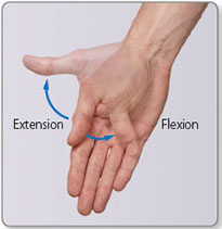
Figure 1.12: Toe dorsiflexion (extension) and plantarflexion (flexion) at the metatarsophalangeal and interphalangeal joints.
zoom view
Figure 1.13: Neck extension and flexion.
11
zoom view
Figure 1.14: Vertebral column extension and flexion.
  • Flexion brings together two surfaces that were originally on the ventral surface of the embryo
  • Extension moves apart two surfaces that were originally on the ventral surface of the embryo
Flexion moves most structures anteriorly whereas extension moves most structures posteriorly. The main exceptions to this occur at the knee, ankle, toes and thumb. For example, plantarflexion is the flexion movement of the feet or toes, dorsiflexion being the extension movement. Flexion of the thumb moves it across the palm in a coronal plane, and extension moves it in the opposite direction.
 
Abduction and adduction
Abduction and adduction occur in the coronal plane (Figures 1.151.20).
  • Abduction moves a structure away from the median plane
  • Adduction moves a structure closer to the median plane
The digits differ in that the movements take place in relation the 3rd digit in the hand and the 2nd digit in the foot. Abduction of the thumb moves it away from the palm in a sagittal plane, and adduction returns it to the anatomical position.12
zoom view
Figure 1.15: Shoulder abduction and adduction.
zoom view
Figure 1.16: Hip abduction and adduction. Hip adduction can take place across the midline if opposite limb is avoided.
13
zoom view
Figure 1.17: Finger (a) adduction and (b) abduction relative to digit 3 (blue line).
zoom view
Figure 1.18: Thumb abduction and adduction.
zoom view
Figure 1.19: Wrist abduction and adduction.
zoom view
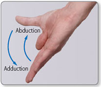
Figure 1.20: Toe (a) abduction and (b) adduction relative to digit 2. The blue line (digit 2) represents the axis of abduction/adduction.
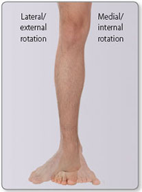
14
zoom view
Figure 1.21: Shoulder rotation: lateral (external) and medial (internal) rotation.
zoom view
Figure 1.22: Hip rotation: medial (internal) and lateral (external) rotation.
zoom view
Figure 1.23: Knee rotation: lateral (external) and medial (internal) rotation.
 
Rotation
Rotation occurs in the transverse plane (Figures 1.211.24).
  • Medial (internal) rotation moves the anterior surface of a limb closer to the median plane
  • Lateral (external) rotation moves the anterior surface of the limb away from the median plane
  • Axial rotation of the vertebral column and atlanto-occipital joints enables turning of the head, neck and trunk to the left or right15
zoom view
Figure 1.24: Axial rotation of the vertebral column.
zoom view
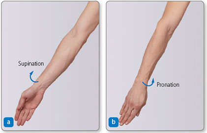
Figure 1.25: Forearm supination and pronation. (a) Supination brings to supine position. (b) Pronation brings forearm to prone position.
 
Pronation and supination
In the forearm:
  • Pronation rotates the radius medially around the ulna such that the palm of the hand faces posteriorly (Figure 1.25)16
    zoom view
    Figure 1.26: Foot pronation (a) and supination (b).
    zoom view
    Figure 1.27: Foot eversion (a) and inversion (b).
  • Supination is the opposite movement: it returns the palm to the anatomical position
In the foot:
  • Pronation lifts its lateral edge such that the sole face away from the median sagittal plane
  • Supination lifts the medial edge such that the sole faces the median sagittal plane (Figure 1.26)
Foot pronation and supination is usually accompanied by foot abduction and adduction, respectively.
 
Inversion and eversion of the foot
  • Inversion is a combination of adduction, supination and plantarflexion
  • Eversion is a combination of abduction, pronation and dorsiflexion (Figure 1.27)17
zoom view
Figure 1.28: Mandibular movements: (a) protrusion ℗ and retrusion ®. (b) mandibular opening.
Both movements occur naturally when attempting to make the sole face medially or laterally with the foot lifted off the floor.
 
Mandibular movements
  • Protrusion moves the mandible (and therefore the chin) anteriorly in a transverse plane
  • Retrusion moves the mandible posteriorly back to the anatomical position (Figure 1.28)
Mandibular opening combines protrusion and rotation.
 
Scapular movements
  • Protraction moves the scapula anterolaterally around the thoracic wall, such as when reaching forward; retraction is the opposite posteromedial movement towards the vertebral column (Figure 1.29)
  • Elevation moves the scapula superiorly on the thoracic wall, such as when shrugging the shoulder; depression is the opposite inferior movement (Figure 1.30)
  • External/lateral rotation makes the glenoid fossa face superolaterally; internal/medial rotation is the opposite movement (Figure 1.31)18
zoom view
Figure 1.29: Scapular movements: (a) retraction and (b) protraction.
zoom view
Figure 1.30: Scapular movements: (a) depression and (b) elevation.
zoom view
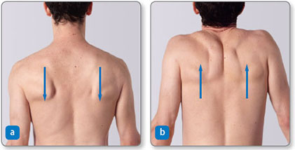
Figure 1.31: Scapular movements: (a) medial (internal) rotation and (b) lateral (external) rotation.
19  
Opposition and reposition of the digits
  • Opposition is the pad-to-pad contact of the thumb and fingers; it involves a specialised movement of the thumb (Figure 1.32) and is essential for fine dexterous activities such as writing and buttoning clothes
  • Reposition returns the thumb to the anatomical position
zoom view
Figure 1.32: Thumb opposition: (a) anterior view and (b) lateral view.
20
zoom view
Figure 1.33: Lateral flexion of the vertebral column: (a) left and (b) right.
zoom view
Figure 1.34: Lateral flexion of the neck to the right.
 
Lateral flexion
Lateral flexion is a lateral movement of the vertebral column in a coronal plane, which results in the head moving away from the midline (median plane) (Figures 1.33 and 1.34).21
zoom view
Figure 1.35: Eye movements. Ⓔ Elevation, Ⓓ depression, Ⓛ left-eye abduction and right-eye adduction, Ⓡ right-eye abduction and left-eye adduction.
zoom view
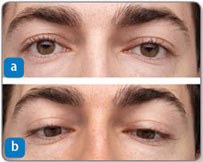
Figure 1.36: Eye movement: convergence. (a) the neutral anatomical position and (b) convergence (with a mild depression)
 
Eye movements
The eyes move around three different mutually perpendicular axes. The movements of abduction–adduction, elevation–depression, and combinations of these are easily observed (Figure 1.35). Most eye movements are conjugate (i.e. both eyes move in the same direction, by the same amount and at the same time), except for convergence, in which both eyes adduct to enable observation of nearby objects (Figure 1.36).22
 
1.3 SENSORY INNERVATION
A knowledge of cutaneous innervation patterns is important when mapping out sensory losses because it provides clues as to the location of damage. Cutaneous innervation is defined by:
  • Dermatomes (Figures 1.37 and 1.38) – the area of skin innervated by a single spinal nerve
  • Cutaneous (named peripheral) nerve areas (Figures 1.39 and 1.40) – the area of skin innervated by a peripheral cutaneous nerve (e.g. ulnar nerve); cutaneous nerve areas may cross parts of one or more dermatomes
As a general rule:
  • Dermatomal sensory losses occur following damage to the spinal nerves or their ventral rami, or the spinal cord
  • Cutaneous nerve area sensory losses occur as a result of nerve lesions in peripheral sites via surgical incisions, laceration or regional compression
  • Superficial cutaneous nerves in the limbs often run close to superficial veins and are therefore vulnerable to injury during cannulation; similarly major peripheral nerves (e.g. femoral nerve) run close to arteries and are vulnerable during arterial access
zoom view
Figure 1.37: An evidence-based pelvic dermatome map. Based on Lee MWL, McPhee RW, Stringer MD. An evidence-based approach to human dermatomes. Clin Anat 2008; 21:363–373.
23
zoom view
Figure 1.38: An evidence-based dermatome map. Based on Lee MWL, McPhee RW, Stringer MD. An evidence-based approach to human dermatomes. Clin Anat 2008; 21:363–373.
24
zoom view
Figure 1.39: Cutaneous innervation of the upper limb.
25
zoom view
Figure 1.40: Cutaneous innervation of the lower limb: (a) anterior view and (b) posterior view.
26