|
|

|
Rheumatic mitral stenosis
|
2 MB
|
|
|
|

|
Rheumatic mitral stenosis
|
2 MB
|
|
|
|

|
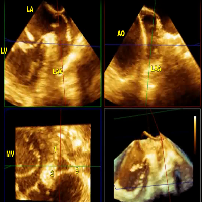
Rheumatic mitral stenosis
|
301 KB
|
|
|
|

|
Rheumatic mitral stenosis
|
1 MB
|
|
|
|

|
Rheumatic mitral stenosis
|
1 MB
|
|
|
|

|
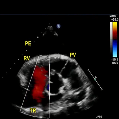
Rheumatic mitral stenosis
|
439 KB
|
|
|
|
|
Rheumatic mitral stenosis
|
2 MB
|
|
|
|

|
Rheumatic mitral stenosis
|
2 MB
|
|
|
|

|
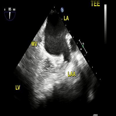
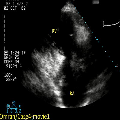
TEE: left atrium enlarged
|
604 KB
|
|
|
|

|
TEE: spontaneous echo contrast in the dilated LA
|
730 KB
|
|
|
|

|
TEE: thrombus in the LAA
|
758 KB
|
|
|
|

|
Mitral valve is only mildly thickened and left atrial appendage is clear
|
474 KB
|
|
|
|

|
Mitral valve thickened and left atrial appendage is clear
|
633 KB
|
|
|
|

|
Mitral valve is only mildly thickened and left atrial appendage is clear
|
219 KB
|
|
|
|

|
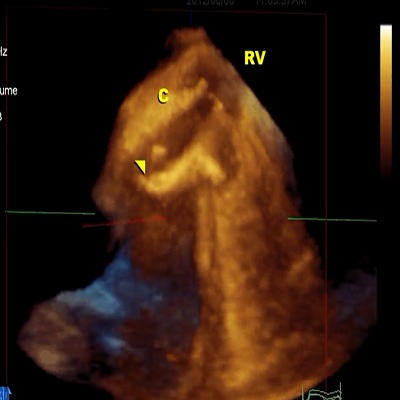
3D TEE: narrow mitral orifice
|
566 KB
|
|
|
|

|
3D TEE: narrow mitral orifice
|
212 KB
|
|
|
|

|
3D TEE: narrow mitral orifice
|
436 KB
|
|
|
|

|
3D TEE: narrow mitral orifice
|
169 KB
|
|
|
|

|
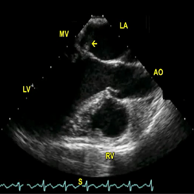
Transthoracic 2D echocardiography apical 4-chamber view
|
3 MB
|
|
|
|

|
Transthoracic 2D echocardiography appendicular view
|
3 MB
|
|
|
|

|
Transesophageal echocardiography mid-esophageal view
|
4 MB
|
|
|
|

|
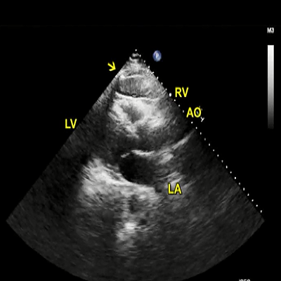
Transthoracic echocardiography off-axis view
|
846 KB
|
|
|
|

|
Transthoracic echocardiography apical 4-chamber view
|
827 KB
|
|
|
|

|
3D TEE zoom mode acquisition of the mitral valve from left atrial perspective
|
2 MB
|
|
|
|

|
TEE zoom mode acquisition of the mitral valve from left ventricular perspective
|
2 MB
|
|
|
|

|
Live 3D TEE zoom mode to guide the interventionist to enter the orifice of the mitral valve
|
9 MB
|
|
|
|

|
Live 3D TEE zoom mode showing full inflation of the Inoue balloon within the mitral orifice
|
11 MB
|
|
|
|

|
Live 3D TEE zoom mode immediately after PMBV
|
3 MB
|
|
|
|

|
Mitral stenosis produced by degenerative calcific disease
|
408 KB
|
|
|
|

|
Mitral stenosis produced by degenerative calcific disease
|
431 KB
|
|
|
|

|
Mitral stenosis produced by degenerative calcific disease
|
487 KB
|
|
|
|

|
Mitral stenosis produced by degenerative calcific disease
|
489 KB
|
|
|
|

|
Mitral valve gradient
|
776 KB
|
|
|
|

|
Mitral valve area
|
822 KB
|
|
|
|

|
High velocity mitral inflow visualized by color Doppler
|
486 KB
|
|
|
|

|
Shadowing (arrow) produced by MV annular calcification
|
955 KB
|
|
|
|

|
Echo: apical four chamber view.
|
1 MB
|
|
|
|

|
Echo: parasternal long axis view.
|
3 MB
|
|
|
|

|
Echo: parasternal short axis view.
|
2 MB
|
|
|
|

|
SSFP HLA (4 = chamber) CMRI
|
314 KB
|
|
|
|

|
SSFP LVOT (3 = chamber) CMRI
|
310 KB
|
|
|
|

|
SSFP SA CMRI
|
319 KB
|
|
|
|

|
MR severity during echocardiographic examination
|
325 KB
|
|
|
|

|
MR severity during echocardiographic examination
|
334 KB
|
|
|
|

|
MR severity during echocardiographic examination
|
328 KB
|
|
|
|

|
MR severity during echocardiographic examination
|
318 KB
|
|
|
|

|
Arrow in point to false tendon
|
937 KB
|
|
|
|

|
Marked prolapse of both MV leaflets (arrow) with severe MR
|
1 MB
|
|
|
|

|
2D TTE
|
1 MB
|
|
|
|

|
2D TTE
|
1 MB
|
|
|
|

|
Preoperative CABG
|
241 KB
|
|
|
|

|
color Doppler study after 6 months: post CABG
|
389 KB
|
|
|
|

|
Apical four chamber view.
|
330 KB
|
|
|
|

|
Vena contracta (VC) area
|
57 KB
|
|
|
|

|
Severity of MR in this patient
|
682 KB
|
|
|
|

|
What does the arrow point to in this adult patient with MR?
|
694 KB
|
|
|
|

|
Flail anterior mitral leaflet
|
8 MB
|
|
|
|

|
Severe posteriorly directed jet of MR
|
8 MB
|
|
|
|

|
3D TEE surgical view of the mitral valve
|
9 MB
|
|
|
|

|
Physio ring annuloplasty
|
2 MB
|
|
|
|

|
Flail middle scallop of posterior mitral leaflet (P2)
|
4 MB
|
|
|
|

|
Severe anteriorly directed jet of MR
|
6 MB
|
|
|
|

|
Multiple ruptured chordae tendineae
|
3 MB
|
|
|
|

|
Severe anteriorly directed jet of mitral regurgitation
|
532 KB
|
|
|
|

|
Flail posterior mitral leaflet
|
156 KB
|
|
|
|

|
Severe mitral regurgitation
|
354 KB
|
|
|
|

|
Flail posterior MV leaflet
|
183 KB
|
|
|
|

|
Redundant MV chordae
|
414 KB
|
|
|
|

|
2D TTE
|
856 KB
|
|
|
|

|
2D TTE
|
807 KB
|
|
|
|

|
2D TTE
|
712 KB
|
|
|
|

|
2D TTE
|
2 MB
|
|
|
|

|
Parasternal long axis views
|
509 KB
|
|
|
|

|
Parasternal long axis views
|
459 KB
|
|
|
|

|
Color Doppler examination aortic valve
|
419 KB
|
|
|
|

|
Aortic stenosis aortic valve area
|
712 KB
|
|
|
|

|
Aortic stenosis aortic valve area
|
1 MB
|
|
|
|

|
AV prolapse
|
646 KB
|
|
|
|

|
Intermittent prolapse of the AV
|
496 KB
|
|
|
|

|
Mild AR
|
489 KB
|
|
|
|

|
2D TTE
|
6 MB
|
|
|
|

|
2D TTE
|
3 MB
|
|
|
|

|
2D TTE
|
3 MB
|
|
|
|

|
Mild aortic regurgitation
|
470 KB
|
|
|
|

|
Mild aortic regurgitation
|
765 KB
|
|
|
|

|
Parasternal long axis view
|
469 KB
|
|
|
|

|
Parasternal long axis view
|
452 KB
|
|
|
|

|
AR PHT
|
682 KB
|
|
|
|

|
Color M-mode echo
|
743 KB
|
|
|
|

|
Mild aortic regurgitation
|
2 MB
|
|
|
|

|
2D TTE
|
439 KB
|
|
|
|
|
2D TTE
|
353 KB
|
|
|
|

|
2D TTE
|
261 KB
|
|
|
|

|
2D TTE
|
165 KB
|
|
|
|

|
MV diastolic flutter
|
178 KB
|
|
|
|

|
Bicuspid AV with equal sized leaflets
|
281 KB
|
|
|
|

|
3D echo: tricuspid morphology
|
397 KB
|
|
|
|

|
2D TTE
|
346 KB
|
|
|
|

|
2D TTE
|
500 KB
|
|
|
|

|
Prominent red signals indicative of backflow
|
194 KB
|
|
|
|

|
3D echo full volume
|
326 KB
|
|
|
|

|
3D MPR mode
|
384 KB
|
|
|
|

|
3D TTE assessment of AR vena contracta
|
312 KB
|
|
|
|

|
Technique of 3D TTE cropping
|
4 MB
|
|
|
|

|
Transthoracic parasternal view
|
115 KB
|
|
|
|

|
Prolapsed leaflet
|
416 KB
|
|
|
|

|
Aortic regurgitation
|
254 KB
|
|
|
|

|
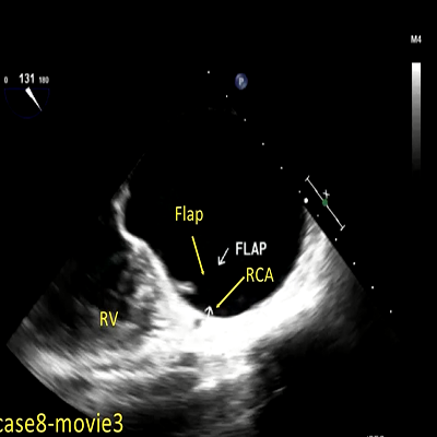
Aortic dissection flap
|
1 MB
|
|
|
|

|
Aneurysmal descending aorta
|
474 KB
|
|
|
|

|
Aneurysmal descending aorta
|
453 KB
|
|
|
|

|
Intimal tear with blood flow
|
1 MB
|
|
|
|

|
Prosthetic reverberations
|
625 KB
|
|
|
|

|
Descending thoracic aorta aneurysm
|
617 KB
|
|
|
|

|
Aortic valve replacement
|
340 KB
|
|
|
|

|
Aortic valve replacement
|
317 KB
|
|
|
|

|
Aortic valve replacement
|
166 KB
|
|
|
|

|
Color Doppler flow signals as compared to the nonperfusing lumen
|
134 KB
|
|
|
|

|
2D TTE
|
329 KB
|
|
|
|

|
Aortic valve replacement
|
129 KB
|
|
|
|

|
Intraoperative TEE in short-axis view of the aortic root
|
2 MB
|
|
|
|

|
TEE short-axis view at the level of aortic annulus
|
2 MB
|
|
|
|
|
TEE in long-axis view dissection flap adjacent
|
2 MB
|
|
|
|

|
Intraoperative immediate postoperative study
|
2 MB
|
|
|
|

|
Transverse plane examination
|
322 KB
|
|
|
|

|
Coronal plane examination
|
327 KB
|
|
|
|

|
Coronal plane examination
|
326 KB
|
|
|
|

|
Coronal plane examination
|
338 KB
|
|
|
|

|
Liver enlargement
|
390 KB
|
|
|
|

|
IVC filter
|
509 KB
|
|
|
|

|
Subcostal/abdominal approaches
|
528 KB
|
|
|
|

|
TR jet entering CS
|
488 KB
|
|
|
|

|
Preoperative 3D TEE, surgical view of the tricuspid valve
|
1 MB
|
|
|
|

|
Preoperative 3D TEE, tricuspid valve from right ventricular aspect
|
1 MB
|
|
|
|

|
3D TEE, tricuspid valve repair
|
2 MB
|
|
|
|

|
Pulmonic valve replacement with a Magna bioprosthetic valve
|
3 MB
|
|
|
|

|
Flail septal TV leaflet
|
532 KB
|
|
|
|

|
Severity of TR in this patient
|
417 KB
|
|
|
|

|
2D TTE
|
629 KB
|
|
|
|

|
2D TTE
|
396 KB
|
|
|
|

|
3D TTE
|
1 MB
|
|
|
|

|
3D TTE
|
1 MB
|
|
|
|

|
3D TTE
|
314 KB
|
|
|
|

|
Tricuspid valve prolapse with mid-to-late systolic regurgitation
|
2 MB
|
|
|
|

|
TTE in tricuspid valve prolapse with mid-to-late systolic regurgitation
|
2 MB
|
|
|
|

|
TTE in tricuspid valve prolapse with mid-to-late systolic regurgitation
|
2 MB
|
|
|
|

|
TTE in tricuspid valve prolapse with mid-to-late systolic regurgitation
|
3 MB
|
|
|
|

|
TTE in tricuspid valve prolapse with mid-to-late systolic regurgitation
|
2 MB
|
|
|
|

|
Pansystolic plus late diastolic tricuspid regurgitation
|
806 KB
|
|
|
|

|
Pansystolic plus late diastolic tricuspid regurgitation
|
710 KB
|
|
|
|

|
CW Doppler velocity waveform with tricuspid regurgitation.
|
519 KB
|
|
|
|

|
Sever tricuspid regurgitation in the patient
|
307 KB
|
|
|
|

|
Pulmonary artery systolic pressure in patient
|
877 KB
|
|
|
|

|
Pulmonary artery systolic pressure in patient
|
715 KB
|
|
|
|

|
Aortic short axis view
|
1022 KB
|
|
|
|

|
Aortic short axis view with long axis of the PA
|
812 KB
|
|
|
|

|
Color Doppler examination of trivial PR
|
1 MB
|
|
|
|

|
The patient is a 74-year-old female with ischemic heart
disease and chronic renal failure. 2D TTE was done
|
416 KB
|
|
|
|

|
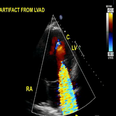
This is an adult patient who underwent left ventricular assist device (LVAD) placement for dilated cardiomyopathy and heart failure.
|
3 MB
|
|
|
|

|
Severity of PR in this patient
|
653 KB
|
|
|
|

|
Mitral valve replacement
|
391 KB
|
|
|
|

|
Mitral valve replacement
|
349 KB
|
|
|
|

|
Prosthetic mitral valve case with fever and left bundle branch block
|
398 KB
|
|
|
|

|
Prosthetic mitral valve case with fever and LBBB
|
299 KB
|
|
|
|

|
Mitral valve replacement
|
916 KB
|
|
|
|

|
2D images of the prosthetic valve
|
242 KB
|
|
|
|

|
3D of the mitral prosthetic valve
|
184 KB
|
|
|
|

|
Transthoracic echocardiographic apical four-chamber view
|
408 KB
|
|
|
|

|
Immobile leaflet of the mitral valve
|
529 KB
|
|
|
|

|
Mobile leaflet and immobile leaflet
|
196 KB
|
|
|
|

|
Mobile leaflet and immobile leaflet
|
137 KB
|
|
|
|

|
Cine-fluoroscopic view of the prosthesis
|
978 KB
|
|
|
|

|
3D transesophageal echocardiography of the prosthetic mitral valve
|
641 KB
|
|
|
|

|
Fluoroscopic of the opening and closing of the prosthetic mitral valve
|
687 KB
|
|
|
|

|
Preoperative 2D TEE in commissural view
|
2 MB
|
|
|
|

|
Severely stenotic bioprosthetic
|
6 MB
|
|
|
|

|
Severely stenotic bioprosthetic mitral valve
|
2 MB
|
|
|
|

|
Severely stenotic bioprosthetic MVR
|
2 MB
|
|
|
|

|
Triple display of MVR
|
2 MB
|
|
|
|

|
LV side, severely stenotic mitral inflow
|
441 KB
|
|
|
|

|
Parasternal long-axis view
|
881 KB
|
|
|
|

|
Parasternal shot-axis view
|
621 KB
|
|
|
|

|
Fluoroscopy of mitral prosthesis
|
1 MB
|
|
|
|

|
Motion of the mitral prosthetic valve
|
1 MB
|
|
|
|

|
Color Doppler image of apical 4-chamber view
|
1 MB
|
|
|
|

|
TEE of 4 chamber view
|
392 KB
|
|
|
|

|
TEE necrotic tissue of the thinned area
|
434 KB
|
|
|
|

|
Prosthetic mitral valve
|
452 KB
|
|
|
|

|
3D TEE with color Doppler
|
125 KB
|
|
|
|

|
Preoperative 2D TEE 4-chamber view
|
6 MB
|
|
|
|

|
Preoperative two-dimensional transthoracic echocardiography
|
509 KB
|
|
|
|

|
color Doppler two large jets of paravalvular leak
|
523 KB
|
|
|
|

|
Re-suturing of the sewing ring
|
581 KB
|
|
|
|

|
Transesophageal echocardiography with color Doppler
|
699 KB
|
|
|
|

|
Three-dimensional confirms evidence of the leaflet perforations
|
587 KB
|
|
|
|

|
Percutaneous plug closure of paravalvular mitral prosthetic regurgitation
|
1 MB
|
|
|
|

|
Percutaneous plug closure of paravalvular mitral prosthetic regurgitation
|
2 MB
|
|
|
|

|
Percutaneous plug closure of paravalvular mitral prosthetic regurgitation
|
3 MB
|
|
|
|

|
Mitral valve replacement
|
1 MB
|
|
|
|

|
Moderate residual MR
|
726 KB
|
|
|
|

|
Moderate residual MR
|
653 KB
|
|
|
|

|
Moderate residual MR
|
382 KB
|
|
|
|

|
Moderate residual MR
|
411 KB
|
|
|
|

|
Preoperative 3D TEE zoom mode acquisition of mitral and tricuspid valve
|
510 KB
|
|
|
|

|
Preoperative 3D TEE of the tricuspid annulus
|
548 KB
|
|
|
|

|
3D TEE full volume color Doppler acquisition of the mitral valve
|
395 KB
|
|
|
|

|
Postoperative 3D TEE after mitral valve repair
|
1 MB
|
|
|
|

|
Mild MR
|
2 MB
|
|
|
|

|
Mild MR
|
1 MB
|
|
|
|

|
MV clip placement
|
1 MB
|
|
|
|

|
TV scallops
|
1 MB
|
|
|
|

|
3D TEE
|
1 MB
|
|
|
|

|
Depicts cropping of the 3D data sets to identify both adjacently placed clips
|
13 MB
|
|
|
|

|
3D multiplanar reconstruction mode
|
10 MB
|
|
|
|

|
Mitral valve clip
|
2 MB
|
|
|
|

|
Mitral valve clip
|
4 MB
|
|
|
|

|
Mitral valve clip
|
3 MB
|
|
|
|

|
Mitral valve clip
|
1 MB
|
|
|
|

|
Mitral valve prolapse
|
343 KB
|
|
|
|

|
Mitral valve prolapse
|
495 KB
|
|
|
|

|
Mitral valve prolapse
|
650 KB
|
|
|
|

|
Mitral valve prolapse
|
440 KB
|
|
|
|

|
Mitral valve prolapse
|
533 KB
|
|
|
|

|
Small ASD with left to right shunt
|
380 KB
|
|
|
|

|
The puncture in the atrial septum was made superior to the PFO region (arrowhead) to provide enough room in the LA to successfully maneuver placement of mitral valve.
|
416 KB
|
|
|
|

|
2D TTE
|
465 KB
|
|
|
|

|
2D TTE
|
466 KB
|
|
|
|

|
Residual MR: color Doppler
|
432 KB
|
|
|
|

|
Anterior and posterior MV leaflets with a typical double orifice MV configuration
|
1 MB
|
|
|
|

|
An 80-year-old male with a murmur of MR. 2D TTE was done
|
1 MB
|
|
|
|

|
Preoperative transthoracic echocardiography in the cath lab
|
807 KB
|
|
|
|

|
Preoperative transthoracic echocardiography with color Doppler
|
444 KB
|
|
|
|

|
Three-dimensional transesophageal echocardiography zoom mode
|
1 MB
|
|
|
|

|
One clip insertion at the medial portion of the mitral closure line
|
1 MB
|
|
|
|

|
Residual mitral regurgitation
|
500 KB
|
|
|
|

|
Residual mitral regurgitation
|
2 MB
|
|
|
|

|
Leaflets and synchronized motion with heartbeats
|
4 MB
|
|
|
|

|
3D TEE zoom mode
|
4 MB
|
|
|
|

|
Fluoroscopic view
|
312 KB
|
|
|
|

|
Fluoroscopic view
|
2 MB
|
|
|
|

|
Preoperative TEE in the cath lab
|
3 MB
|
|
|
|

|
Postoperative TEE in the cath lab
|
1 MB
|
|
|
|

|
3D TEE zoom mode acquisition of the mitral valve from LV side
|
2 MB
|
|
|
|

|
2D TEE 10 minutes after insertion of the clip increased MR
|
4 MB
|
|
|
|

|
3D TEE confirmation of partial detachment of the clip
|
2 MB
|
|
|
|

|
Fluoroscopy view of the clip showing jerky movements
|
2 MB
|
|
|
|

|
2D TEE in the cath lab
|
2 MB
|
|
|
|

|
2D TEE on day 6 after procedure
|
1 MB
|
|
|
|

|
2D TEE for better visualization
|
1 MB
|
|
|
|

|
2D TEE in 4-chamber view
|
1 MB
|
|
|
|

|
Normal leaflet motion by 2D echo
|
625 KB
|
|
|
|

|
2 DTEE: flail bioprosthetic AVR with severe AR
|
619 KB
|
|
|
|

|
2DTEE: flail AVR with thickened leaflets
|
1 MB
|
|
|
|

|
2DTEE (left) and 3DTEE (right): flail AVR with thickened leaflets
|
1 MB
|
|
|
|

|
TTE parasternal long axis view rocking motion of the aortic bioprosthesis
|
1 MB
|
|
|
|

|
Transthoracic echocardiography apical long axis view
|
1 MB
|
|
|
|

|
TTE apical long axis view with color Doppler demonstrates paravalvular leak
|
1 MB
|
|
|
|

|
TEE midesophageal long-axis view
|
527 KB
|
|
|
|

|
TEE midesophageal long-axis view: color Doppler
|
368 KB
|
|
|
|

|
TEE transgastric long-axis view (120°)
|
552 KB
|
|
|
|

|
Intraoperative 3D TEE in cath lab during TAVR
|
4 MB
|
|
|
|

|
2D TEE in the cath lab in long-axis view
|
3 MB
|
|
|
|

|
3D TEE zoom mode acquisition
|
3 MB
|
|
|
|

|
3D TEE in cath lab one year after TAVR
|
1 MB
|
|
|
|

|
Zoom mode live acquisition
|
13 MB
|
|
|
|

|
Preoperative 2D TEE in long-axis view
|
4 MB
|
|
|
|

|
Preoperative 2D TEE in long-axis view: bioprosthetic aortic valve
|
4 MB
|
|
|
|

|
Color flow signals inside the cavities
|
4 MB
|
|
|
|

|
3D TEE in long-axis view
|
857 KB
|
|
|
|

|
3D TEE full volume acquisition of the aortic root
|
736 KB
|
|
|
|

|
Vegetation and extension of the infection to the aortic-mitral curtain
|
808 KB
|
|
|
|

|
Restricted motion of two posterior cusps of the bioprosthetic mitral valve
|
1 MB
|
|
|
|

|
Severe transvalvular mitral regurgitation
|
462 KB
|
|
|
|

|
Mitral cusps with normally functioning bioprosthetic mitral valve
|
1 MB
|
|
|
|

|
Full volume color Doppler coaptation of the leaflets
|
417 KB
|
|
|
|

|
Two-dimensional transesophageal echocardiography
|
2 MB
|
|
|
|

|
Two-dimensional transesophageal echocardiography
|
2 MB
|
|
|
|

|
Parasternal long and short axis views with Doppler color flow
|
16 MB
|
|
|
|

|
Aortic root abscess around a bioprosthetic aortic valve forming a fistulous communication to left atrium
|
13 MB
|
|
|
|

|
TEE color Doppler shows mitral regurgitation.
|
175 KB
|
|
|
|

|
TEE color Doppler shows mitral regurgitation.
|
322 KB
|
|
|
|

|
In this video TEE long axis view showing AVR.
|
319 KB
|
|
|
|

|
TEE color Doppler
|
494 KB
|
|
|
|

|
TEE color Doppler
|
305 KB
|
|
|
|

|
3D TEE surgeon’s view of the mitral valve with anterior (A1, A2 and A3) and posterior (P1, P2 and P3) scallops
|
2 MB
|
|
|
|

|
3D TEE: surgical view of the mitral valve
|
627 KB
|
|
|
|

|
3D TEE in mitral commissural
|
508 KB
|
|
|
|

|
3D TEE surgical view of the mitral valve
|
535 KB
|
|
|
|

|
Intraoperative 3D TEE mitral valve after repair
|
601 KB
|
|
|
|

|
Intraoperative 3D TEE : no residual mitral regurgitation
|
358 KB
|
|
|
|
|
TTE parasternal long axis view revealed a large mobile echo density mass
|
372 KB
|
|
|
|

|
TTE parasternal long axis view and color Doppler revealed a perforation/rupture of the anterior leaflet of the mitral valve with torrential MR
|
370 KB
|
|
|
|

|
TTE apical four chamber view zoomed at the mitral valve
|
971 KB
|
|
|
|

|
TTE apical five chamber view: perforation of the anterior mitral leaflet
|
923 KB
|
|
|
|

|
Parasternal long axis view
|
1 MB
|
|
|
|

|
Color Doppler examination: severe MR
|
1 MB
|
|
|
|

|
Color Doppler examination: severe MR
|
462 KB
|
|
|
|

|
Color Doppler examination: severe MR
|
470 KB
|
|
|
|

|
Anterior MV leaflet
|
952 KB
|
|
|
|

|
Color Doppler: severe MR
|
360 KB
|
|
|
|

|
3D TTE: anterior MV leaflet
|
627 KB
|
|
|
|

|
3DTTE: measurement of the area of vena contracta
|
569 KB
|
|
|
|

|
A four chamber view revealed an echo density attached to the tip of the posterior leaflet of the mitral valve involving both the atrial and ventricular sides of the valve.
|
296 KB
|
|
|
|

|
Color Doppler: jet of mitral regurgitation.
|
260 KB
|
|
|
|

|
Parasternal long axis
|
751 KB
|
|
|
|

|
Basal short axis
|
402 KB
|
|
|
|

|
LV short axis
|
757 KB
|
|
|
|

|
Apical 5 chamber view
|
268 KB
|
|
|
|

|
Apical 4 chamber view
|
774 KB
|
|
|
|

|
Apical 2 chamber view
|
753 KB
|
|
|
|

|
Transthoracic echocardiography
|
509 KB
|
|
|
|

|
Transthoracic echocardiography
|
514 KB
|
|
|
|

|
Transthoracic echocardiography
|
261 KB
|
|
|
|

|
Transthoracic echocardiography
|
249 KB
|
|
|
|

|
Transesophageal echocardiography
|
265 KB
|
|
|
|

|
Transesophageal echocardiography
|
763 KB
|
|
|
|

|
Transesophageal echocardiography
|
240 KB
|
|
|
|

|
Transesophageal echocardiography
|
263 KB
|
|
|
|

|
Transesophageal echocardiography
|
258 KB
|
|
|
|

|
Transesophageal echocardiography
|
256 KB
|
|
|
|

|
3D echocardiography
|
167 KB
|
|
|
|

|
3D echocardiography
|
234 KB
|
|
|
|

|
3D echocardiography
|
206 KB
|
|
|
|

|
3D echocardiography
|
200 KB
|
|
|
|

|
3D echocardiography
|
206 KB
|
|
|
|

|
3D echocardiography
|
244 KB
|
|
|
|

|
TEE view of RA, LA, AV, RV
|
770 KB
|
|
|
|

|
TEE view of right atrium
|
779 KB
|
|
|
|

|
TEE view of CATH
|
752 KB
|
|
|
|

|
Two-dimensional transesophageal echocardiography: intracardiac defibrillator
|
207 KB
|
|
|
|

|
Two-dimensional transesophageal echocardiography: intracardiac defibrillator
|
210 KB
|
|
|
|

|
Two-dimensional transesophageal echocardiography: intracardiac defibrillator
|
203 KB
|
|
|
|

|
3D echocardiography
|
169 KB
|
|
|
|

|
Subcostal examination
|
721 KB
|
|
|
|

|
Bicuspid AV with mild thickening
|
1 MB
|
|
|
|

|
PE: thickened TV
|
543 KB
|
|
|
|

|
Large PE
|
950 KB
|
|
|
|
|
Large PE. Mild TR
|
915 KB
|
|
|
|

|
Spontaneous contrast in IVC
|
1000 KB
|
|
|
|

|
Large pericardial thrombus
|
997 KB
|
|
|
|

|
Catheter in the right heart
|
866 KB
|
|
|
|

|
LVO-RA shunt (S) just beneath the TV
|
766 KB
|
|
|
|

|
Vegetations on both atrial and ventricular aspects of TV
|
900 KB
|
|
|
|

|
Vegetations on both atrial and ventricular aspects of TV
|
1000 KB
|
|
|
|

|
LV-RV and LV-RA shunts
|
1 MB
|
|
|
|

|
LV-RA/RV shunt (Gerbode shunt) visualized subcostally
|
2 MB
|
|
|
|

|
2D TTE in tetralogy of Fallot postrepair
|
454 KB
|
|
|
|

|
Peak and mean gradients across the TV
|
413 KB
|
|
|
|

|
Peak and mean gradients across the TV
|
457 KB
|
|
|
|

|
Peak and mean gradients across the tricuspid valve of 77 mm Hg
|
353 KB
|
|
|
|

|
Multiplane 2D TEE in tetralogy of Fallot postrepair
|
1 MB
|
|
|
|

|
2D TTE in tetralogy of Fallot postrepair.
|
389 KB
|
|
|
|

|
Torrential conduit valve regurgitation.
|
469 KB
|
|
|
|

|
3D TTE in tetralogy of Fallot postrepair
|
372 KB
|
|
|
|

|
3D TTE in tetralogy of Fallot postrepair
|
499 KB
|
|
|
|

|
3D TTE in tetralogy of Fallot postrepair
|
473 KB
|
|
|
|

|
3D TTE in tetralogy of Fallot postrepair
|
530 KB
|
|
|
|

|
3D TTE in tetralogy of Fallot postrepair
|
288 KB
|
|
|
|
|
3D TTE in tetralogy of Fallot postrepair
|
402 KB
|
|
|
|

|
3D TTE in tetralogy of Fallot postrepair
|
167 KB
|
|
|
|

|
Volume (V1) of the largest vegetation
|
352 KB
|
|
|
|

|
2D TTE
|
405 KB
|
|
|
|

|
2D TTE parasternal long axis view
|
555 KB
|
|
|
|

|
2D TTE parasternal short axis view
|
631 KB
|
|
|
|

|
Hypokinetic apical inferoseptum
|
1 MB
|
|
|
|

|
Apical RV free wall hypokinesis
|
1 MB
|
|
|
|

|
Right ventricle apical dyskinesis
|
490 KB
|
|
|
|

|
2D TTE parasternal long-axis view
|
234 KB
|
|
|
|

|
Left ventricle infero-lateral wall dyskinesis
|
1 MB
|
|
|
|

|
2D TTE: apical 2 chamber view
|
554 KB
|
|
|
|

|
Aneurysmal proximal LV inferior wall
|
876 KB
|
|
|
|

|
LV apical aneurysm
|
1 MB
|
|
|
|

|
LV apical aneurysm
|
1 MB
|
|
|
|

|
LV apical aneurysm
|
906 KB
|
|
|
|

|
LV apical aneurysm
|
1 MB
|
|
|
|

|
Large echo density in the LV apex
|
1 MB
|
|
|
|

|
Large echo density in the LV apex
|
803 KB
|
|
|
|

|
Large echo density in the LV apex
|
1 MB
|
|
|
|

|
Transthoracic apical view
|
899 KB
|
|
|
|

|
Transesophageal echocardiography
|
465 KB
|
|
|
|

|
LV angiogram
|
1 MB
|
|
|
|

|
Cardiac MRI
|
529 KB
|
|
|
|

|
Cardiac MRI
|
421 KB
|
|
|
|

|
Multiple pseudoaneurysms and pleural effusion
|
1 MB
|
|
|
|

|
Multiple pseudoaneurysms and pleural effusion
|
1 MB
|
|
|
|

|
Multiple pseudoaneurysms and pleural effusion
|
627 KB
|
|
|
|

|
Ventricular septal rupture with left to right shunt
|
3 MB
|
|
|
|

|
Ventricular septal rupture with left to right shunt
|
3 MB
|
|
|
|

|
En-face view of the VSD
|
459 KB
|
|
|
|

|
Postoperative MI VSD
|
472 KB
|
|
|
|

|
Postoperative myocardial infarction VSD
|
431 KB
|
|
|
|

|
Postoperative patch exclusion of VSD
|
396 KB
|
|
|
|

|
Ischemic mitral valve with restricted posterior leaflet
|
939 KB
|
|
|
|

|
TEE: posterior direction of mitral regurgitation
|
999 KB
|
|
|
|

|
TEE: posterior leaflet (PL) of mitral valve
|
1011 KB
|
|
|
|

|
TEE: severe mitral regurgitation
|
179 KB
|
|
|
|

|
TEE: transgastric view
|
174 KB
|
|
|
|

|
TEE: transgastric view
|
197 KB
|
|
|
|
|
Ruptured papillary muscle
|
1 MB
|
|
|
|

|
Ruptured papillary muscle
|
1 MB
|
|
|
|

|
Mitral regurgitation
|
3 MB
|
|
|
|

|
Pericardial effusion
|
1 MB
|
|
|
|

|
Resting LVOT velocity
|
1 MB
|
|
|
|

|
LVOT interrogation
|
373 KB
|
|
|
|

|
Systolic anterior motion of the mitral valve typical of hypertrophic cardiomyopathy
|
492 KB
|
|
|
|

|
High left ventricular outflow tract gradient show apical 4 views
|
204 KB
|
|
|
|

|
High left ventricular outflow tract gradient show 2 chamber views
|
188 KB
|
|
|
|

|
Proximal left main (LMCA) and right (RCA) coronary arteries
|
872 KB
|
|
|
|

|
Artifacts mimicking small plaques in LMCA
|
1 MB
|
|
|
|

|
2D TTE: parasternal long-axis view
|
629 KB
|
|
|
|

|
2D TTE: Parasternal long-axis
|
1 MB
|
|
|
|

|
Three-dimensional transthoracic echocardiography
|
629 KB
|
|
|
|

|
Three-dimensional transthoracic echocardiography
|
261 KB
|
|
|
|

|
Pericardial/epicardial fat pad
|
972 KB
|
|
|
|

|
Pericardial/epicardial fat pad
|
961 KB
|
|
|
|
|
Pericardial/epicardial fat pad
|
886 KB
|
|
|
|

|
IVCs: subcostal view
|
474 KB
|
|
|
|

|
IVCs: subcostal view
|
1 MB
|
|
|
|

|
IVCs: subcostal view
|
618 KB
|
|
|
|

|
IVCs: subcostal view
|
631 KB
|
|
|
|

|
IVCs: subcostal view
|
354 KB
|
|
|
|

|
Color M-mode examination
|
702 KB
|
|
|
|

|
Aneurysm
|
364 KB
|
|
|
|

|
Right ventricular apical aneurysm
|
376 KB
|
|
|
|

|
Right ventricular apical aneurysm
|
302 KB
|
|
|
|

|
Right ventricular apical aneurysm
|
443 KB
|
|
|
|

|
Right ventricular apical aneurysm
|
1 MB
|
|
|
|

|
Right ventricular apical aneurysm
|
1 MB
|
|
|
|

|
Left ventricle extremely poor function
|
423 KB
|
|
|
|

|
Left ventricle extremely poor function
|
398 KB
|
|
|
|

|
Trabeculations in the LV apex
|
400 KB
|
|
|
|

|
Trabeculations in the LV apex
|
429 KB
|
|
|
|

|
Left atrial appendage (LAA) is clear without a clot
|
410 KB
|
|
|
|

|
Apical long axis view
|
390 KB
|
|
|
|

|
Parasternal long axis view
|
1 MB
|
|
|
|

|
Apical 2-chamber view
|
975 KB
|
|
|
|

|
Left ventricle endocardial border
|
1 MB
|
|
|
|

|
Left ventricle endocardial border
|
1 MB
|
|
|
|

|
Left ventricle endocardial border
|
830 KB
|
|
|
|

|
Large mobile thrombus in the LV apex
|
1 MB
|
|
|
|

|
Large mobile thrombus in the LV apex
|
2 MB
|
|
|
|

|
Apical 4-chamber view
|
855 KB
|
|
|
|

|
Small associated trabeculations
|
1 MB
|
|
|
|

|
Apical 4 chamber and parasternal long axis
|
370 KB
|
|
|
|

|
Apical 4 chamber and parasternal long axis
|
354 KB
|
|
|
|

|
Apical 4 chamber and parasternal long axis
|
373 KB
|
|
|
|

|
Apical 4 chamber and parasternal long axis
|
317 KB
|
|
|
|

|
Both left ventricle and right ventricle function compared
|
823 KB
|
|
|
|

|
Both left ventricle and right ventricle function compared
|
814 KB
|
|
|
|

|
Both left ventricle and right ventricle function compared
|
1 MB
|
|
|
|

|
Both left ventricle and right ventricle function compared
|
1 MB
|
|
|
|

|
Both left ventricle and right ventricle function compared
|
764 KB
|
|
|
|

|
Parasternal long axis view
|
1 MB
|
|
|
|

|
Parasternal short axis view
|
1 MB
|
|
|
|

|
Apical 4-chamber view
|
443 KB
|
|
|
|

|
Apical 2-chamber view
|
461 KB
|
|
|
|

|
Apical 5-chamber view
|
437 KB
|
|
|
|

|
2D echoes before left panel and after right panel
|
607 KB
|
|
|
|

|
3D echo
|
577 KB
|
|
|
|

|
Transthoracic echo
|
159 KB
|
|
|
|

|
Global longitudinal Strain (Bulls Eye) by 3D echo
|
196 KB
|
|
|
|

|
pacing site, quadripolar, aortic valve,
|
159 KB
|
|
|
|

|
Tissue synchronous imaging (TSI)
|
545 KB
|
|
|
|

|
Three-dimensional transthoracic echocardiogram
|
599 KB
|
|
|
|

|
3D-TTE: inflow catheter tip
|
582 KB
|
|
|
|

|
Parasternal long axis view
|
502 KB
|
|
|
|

|
Parasternal long axis view
|
401 KB
|
|
|
|

|
Occasional partial opening and closing of the AV
|
538 KB
|
|
|
|

|
AV short axis view
|
375 KB
|
|
|
|

|
LV short axis view at the level of the papillary muscle
|
297 KB
|
|
|
|

|
Short axis view near the level of LV apex
|
206 KB
|
|
|
|

|
Red flow signals representing LV blood
|
295 KB
|
|
|
|
|
color Doppler artifact
|
129 KB
|
|
|
|

|
M-mode LVAD
|
603 KB
|
|
|
|

|
Adult patient who underwent mitral valve replacement (MVR) and left ventricular assist device.
|
1 MB
|
|
|
|

|
Aortic arch and descending aorta
|
428 KB
|
|
|
|

|
PW Doppler examination
|
161 KB
|
|
|
|

|
Apical thrombus
|
449 KB
|
|
|
|

|
Apical thrombus
|
392 KB
|
|
|
|

|
Transthoracic echocardiogram (TTE)
|
376 KB
|
|
|
|

|
Left ventricle (LV) angiogram
|
4 MB
|
|
|
|

|
TTE apical 4-chamber
|
635 KB
|
|
|
|

|
2D TTE five-chamber views
|
186 KB
|
|
|
|

|
2D TTE five-chamber views with color Doppler
|
191 KB
|
|
|
|

|
2D TTE
|
355 KB
|
|
|
|

|
3D TTE
|
81 KB
|
|
|
|

|
3D TTE
|
130 KB
|
|
|
|

|
Two-dimensional transthoracic echocardiography
|
1 MB
|
|
|
|

|
Echo contrast study
|
836 KB
|
|
|
|

|
Echo contrast study
|
391 KB
|
|
|
|

|
Echo contrast study
|
757 KB
|
|
|
|

|
Short-axis view of non-compaction cardiomyopathy
|
328 KB
|
|
|
|

|
Short-axis view of non-compaction cardiomyopathy
|
348 KB
|
|
|
|

|
Four-chamber view of contrast echocardiogram
|
347 KB
|
|
|
|

|
Parasternal long axis view
|
214 KB
|
|
|
|

|
Patient with suspected amyloidosis
|
1 MB
|
|
|
|

|
Patient with suspected amyloidosis
|
1 MB
|
|
|
|

|
Patient with suspected amyloidosis
|
989 KB
|
|
|
|

|
2D parasternal long axis views of LV
|
316 KB
|
|
|
|

|
2D parasternal short axis views of LV
|
316 KB
|
|
|
|

|
Hypertrophied LV and RV walls
|
987 KB
|
|
|
|

|
Hypertrophied LV and RV walls
|
673 KB
|
|
|
|

|
Thickened RV walls
|
760 KB
|
|
|
|

|
Thickened right ventricular walls
|
1023 KB
|
|
|
|

|
Parasternal long-axis view
|
1 MB
|
|
|
|

|
Apical 4-chamber view: pacemaker is visualized in the right heart
|
1 MB
|
|
|
|

|
Parasternal long-axis view
|
1 MB
|
|
|
|

|
Apical 4 chamber view
|
1 MB
|
|
|
|

|
LV longitudinal strain
|
284 KB
|
|
|
|

|
Parasternal long-axis view
|
1 MB
|
|
|
|

|
Apical 4 chamber view
|
1 MB
|
|
|
|

|
Parasternal long-axis view
|
1 MB
|
|
|
|

|
Apical 4 chamber view
|
1 MB
|
|
|
|

|
LV longitudinal strain
|
259 KB
|
|
|
|

|
Left ventricular longitudinal strain
|
244 KB
|
|
|
|

|
Echocardiographic cine loops from this patient: apical 4-chamber TTE view
|
809 KB
|
|
|
|

|
Echocardiographic cine loops from this patient: parasternal short axis TTE view
|
782 KB
|
|
|
|

|
Echocardiographic cine loops from this patient: midesophageal three chamber MRI view
|
768 KB
|
|
|
|

|
Cardiac MRI cine loops from this patient: four chamber MRI view.
|
193 KB
|
|
|
|

|
Cardiac MRI cine loops from this patient: two chamber MRI view
|
189 KB
|
|
|
|

|
Cardiac MRI cine loops from this patient: three chamber MRI view.
|
185 KB
|
|
|
|

|
2D TTE
|
622 KB
|
|
|
|

|
Severe tricuspid regurgitation
|
981 KB
|
|
|
|

|
Severe pulmonary regurgitation (arrow) by color Doppler
|
356 KB
|
|
|
|

|
Apical 4 chamber and subcostal views in an adult patient
|
676 KB
|
|
|
|

|
Apical 4 chamber and subcostal views in an adult patient
|
406 KB
|
|
|
|

|
Apical 4 chamber and subcostal views in an adult patient
|
479 KB
|
|
|
|

|
Maximum (MAX) resting gradient (GRD) of 27 mm Hg across the LVOT
|
835 KB
|
|
|
|

|
Maximum (MAX) resting gradient (GRD) of 27 mm Hg across the LVOT increased to 67 mm Hg with the Valsalva maneuver.
|
4 MB
|
|
|
|

|
LV apical aneurysm (AN)
|
651 KB
|
|
|
|

|
LV apical aneurysm (AN)
|
935 KB
|
|
|
|

|
LV apical aneurysm (AN)
|
847 KB
|
|
|
|

|
3D TTE in hypertrophic cardiomyopathy
|
222 KB
|
|
|
|

|
3D TTE in hypertrophic cardiomyopathy
|
9 MB
|
|
|
|

|
3D TTE in hypertrophic cardiomyopathy
|
4 MB
|
|
|
|

|
3D TTE in hypertrophic cardiomyopathy
|
378 KB
|
|
|
|
|
Left ventriculogram
|
1 MB
|
|
|
|

|
Apical 4-chamber view
|
584 KB
|
|
|
|

|
Apical 2-chamber view
|
519 KB
|
|
|
|

|
Multiple trabeculations in LV apex
|
432 KB
|
|
|
|

|
Multiple trabeculations in LV apex
|
496 KB
|
|
|
|

|
Multiple trabeculations in LV apex
|
503 KB
|
|
|
|

|
Multiple trabeculations in LV apex
|
1 MB
|
|
|
|

|
Multiple trabeculations in LV apex
|
138 KB
|
|
|
|

|
Apical hypertrophic obstructive cardiomyopathy
|
2 MB
|
|
|
|

|
Apical hypertrophic obstructive cardiomyopathy
|
1 MB
|
|
|
|

|
Apical hypertrophic obstructive cardiomyopathy
|
2 MB
|
|
|
|

|
Apical hypertrophic obstructive cardiomyopathy
|
2 MB
|
|
|
|

|
Apical hypertrophic obstructive cardiomyopathy
|
2 MB
|
|
|
|

|
Apical hypertrophic obstructive cardiomyopathy
|
549 KB
|
|
|
|

|
Apical hypertrophic obstructive cardiomyopathy
|
395 KB
|
|
|
|

|
Apical hypertrophic obstructive cardiomyopathy
|
322 KB
|
|
|
|

|
Apical hypertrophic obstructive cardiomyopathy
|
343 KB
|
|
|
|

|
Two-dimensional transthoracic echocardiography. Arrow points to mitral annulus calcification and MR.
|
2 MB
|
|
|
|

|
Two-dimensional transthoracic echocardiography
|
2 MB
|
|
|
|

|
Two-dimensional transthoracic echocardiography
|
2 MB
|
|
|
|

|
Two-dimensional transthoracic echocardiography. Arrowheads demonstrate LV subendocardial calcification and calcification of basal RV free wall.
|
2 MB
|
|
|
|

|
Three-dimensional transthoracic echocardiography
|
13 MB
|
|
|
|

|
Three-dimensional transthoracic echocardiography
|
18 MB
|
|
|
|

|
Three-dimensional transthoracic echocardiography
|
17 MB
|
|
|
|

|
Three-dimensional transthoracic echocardiography
|
2 MB
|
|
|
|

|
Three-dimensional transthoracic echocardiography
|
12 MB
|
|
|
|

|
Reverberation artifact from MV
|
781 KB
|
|
|
|

|
#1 represents the transplanted LA and #2 a remnant of old LA.
|
800 KB
|
|
|
|

|
#1 represents the transplanted LA and #2 a remnant of old LA.
|
815 KB
|
|
|
|

|
Poor RV function with decreased TAPSE
|
3 MB
|
|
|
|

|
Right atrial inversion
|
158 KB
|
|
|
|
|
Cardiac transplantation. Apical 5 chamber view
|
1 MB
|
|
|
|

|
Parasternal long-axis views are shown from an adult
|
793 KB
|
|
|
|

|
Color flow signals can be visualized moving from the vein into LA
|
726 KB
|
|
|
|

|
Parasternal long-axis view from an adult patient
|
832 KB
|
|
|
|

|
2D echocardiograhy. Short axis view. Arrow shows space(S), adjacent to left ventricle (LV).
|
4 MB
|
|
|
|

|
2D TTE. Short axis view. Arrow shows space(S), adjacent to left ventricle (LV).
|
5 MB
|
|
|
|

|
2D echocardiograhy. Short axis view. Arrow shows space(S), adjacent to left ventricle (LV).
|
5 MB
|
|
|
|

|
Parasternal long-axis view shows thickening of the posterior pericardium.
|
3 MB
|
|
|
|

|
Two-dimensional transthoracic echocardiography
|
377 KB
|
|
|
|

|
2D TTE: Thickened anterior and posterior pericardium with no obvious constriction
|
388 KB
|
|
|
|

|
Two-dimensional echocardiography with an apical 4-chamber view
|
773 KB
|
|
|
|

|
Parasternal short-axis view
|
704 KB
|
|
|
|

|
2D echocardiogram
|
370 KB
|
|
|
|

|
2D echocardiogram
|
373 KB
|
|
|
|

|
Apical 4 chamber view
|
498 KB
|
|
|
|

|
Parasternal short axis view
|
503 KB
|
|
|
|

|
Inferior vena cava
|
639 KB
|
|
|
|

|
Preoperative TTE in short-axis view showing ventricular septal bounce.
|
4 MB
|
|
|
|

|
Preoperative TTE showing dilated noncollapsing IVC (IVC plethora)
|
3 MB
|
|
|
|

|
Follow-up TTE after 2 years showing normal motion of the ventricular septum (no septal bounce).
|
1 MB
|
|
|
|

|
Follow-up TTE after 2 years showing small size IVC with good respiratory collapse.
|
5 MB
|
|
|
|

|
Preoperative assessment of left ventricular global circumferential strain (GCS) at the basal LV = –18.5% which is lower than normal.
|
821 KB
|
|
|
|

|
Postoperative basal LV strain, GCS = –21% which is in the normal range.
|
653 KB
|
|
|
|

|
Preoperative assessment of left ventricular global circumferential strain (GCS) at the apical level = –15.1% which is severely reduced.
|
811 KB
|
|
|
|

|
Postoperative GCS at apical LV = –55% which is super normal.
|
637 KB
|
|
|
|

|
Preoperative assessment of left ventricular global longitudinal strain (GLS) is –16.4% which is slightly below normal.
|
574 KB
|
|
|
|

|
Postoperative GLS = –18% which is in the normal range.
|
683 KB
|
|
|
|

|
Examination performed from the left and right back in this patient
|
449 KB
|
|
|
|

|
Examination performed from the left and right back in this patient
|
343 KB
|
|
|
|

|
Examination performed from the left and right back in this patient
|
863 KB
|
|
|
|

|
The large echo free space behind the LV in the parasternal long-axis view clearly extends beyond the descending AO typical of left PLE.
|
308 KB
|
|
|
|

|
No echo free space is noted subcostally.
|
463 KB
|
|
|
|

|
Arrows point to show fibrin strands in PLE.
|
333 KB
|
|
|
|
|
Arrow show falciform ligament
|
372 KB
|
|
|
|

|
Arrow show falciform ligament
|
386 KB
|
|
|
|

|
Subcostal examination. Arrow points to ascites and the arrowhead to falciform ligament
|
553 KB
|
|
|
|

|
Bubble study is positive for intrapulmonary shunting.
|
4 MB
|
|
|
|

|
Echo free space due to ascites
|
71 KB
|
|
|
|

|
Visualization of coils of intestine (arrows) during abdominal examination provides a clue that the fluid (arrow head) collection is ascites.
|
71 KB
|
|
|
|

|
This video clip visualization of coils of intestine (arrows) during abdominal examination provides a clue that the fluid (arrow head) collection is ascites.
|
78 KB
|
|
|
|

|
Presence of pericardial effusion (PE) behind left ventricle and left atrium (arrow)
|
583 KB
|
|
|
|

|
Dilated coronary sinus
|
618 KB
|
|
|
|

|
Normal LV function
|
744 KB
|
|
|
|

|
Presence of ascites (transverse arrow) with falciform ligament
|
700 KB
|
|
|
|

|
Echo free space is produced by breast implants.
|
381 KB
|
|
|
|

|
Echo free space is produced by breast implants.
|
391 KB
|
|
|
|

|
Echo free space is produced by breast implants.
|
1 MB
|
|
|
|

|
Echo free space is produced by breast implants.
|
232 KB
|
|
|
|

|
TTE: subcostal view
|
3 MB
|
|
|
|

|
TTE: apical four chamber view
|
1 MB
|
|
|
|

|
Transthoracic apical view
|
520 KB
|
|
|
|

|
Transesophageal echocardiography
|
769 KB
|
|
|
|

|
Transesophageal echocardiography
|
759 KB
|
|
|
|

|
Transesophageal echocardiography
|
1 MB
|
|
|
|

|
Transesophageal echocardiography
|
367 KB
|
|
|
|

|
Transesophageal echocardiography
|
367 KB
|
|
|
|

|
Post catheter removal
|
1 MB
|
|
|
|

|
3D TEE
|
4 MB
|
|
|
|

|
3D TEE
|
3 MB
|
|
|
|

|
Two-dimensional transthoracic echocardiography
|
372 KB
|
|
|
|

|
Two-dimensional transthoracic echocardiography
|
195 KB
|
|
|
|

|
Two-dimensional transthoracic echocardiography
|
176 KB
|
|
|
|

|
Two-dimensional transthoracic echocardiography
|
359 KB
|
|
|
|

|
Two-dimensional transthoracic echocardiography
|
355 KB
|
|
|
|

|
Three-dimensional transthoracic echocardiography
|
72 KB
|
|
|
|

|
Three-dimensional transthoracic echocardiography
|
71 KB
|
|
|
|

|
Three-dimensional transthoracic echocardiography
|
88 KB
|
|
|
|

|
Apical 4 chamber
|
521 KB
|
|
|
|

|
Apical 4 chamber zoomed
|
520 KB
|
|
|
|

|
Subcostal view, IVC
|
510 KB
|
|
|
|

|
2D echocardiography
|
340 KB
|
|
|
|

|
Echocardiography IVC view
|
337 KB
|
|
|
|

|
Apical 4 chamber with ultrasound contrast. Arrow points to bubbles appearing in the LV
|
302 KB
|
|
|
|

|
Subcostal/IVC view with ultrasound contrast with delayed filling of the IVC. Arrow shows appearance of bubbles in the IVC
|
332 KB
|
|
|
|

|
Presence of mass or thrombus occluding the inferior vena cava (revealing as contrast defect)
|
1 MB
|
|
|
|

|
Transthoracic parasternal long axis view
|
403 KB
|
|
|
|

|
Transthoracic apical 4 chamber view
|
397 KB
|
|
|
|

|
2D TEE
|
2 MB
|
|
|
|

|
3D TEE
|
1 MB
|
|
|
|

|
3D TEE
|
2 MB
|
|
|
|

|
Two-dimensional transesophageal echocardiography of a thrombus in transit through a patent foramen ovale
|
2 MB
|
|
|
|

|
2D TEE of a thrombus in transit through a patent foramen ovale
|
2 MB
|
|
|
|

|
Two-dimensional transesophageal echo of thrombus in transit through a patent foramen ovale
|
1 MB
|
|
|
|

|
3D TEE
|
1 MB
|
|
|
|

|
3D TEE
|
1 MB
|
|
|
|

|
3D TEE
|
1 MB
|
|
|
|

|
3D TEE
|
1 MB
|
|
|
|

|
3D TEE
|
3 MB
|
|
|
|

|
3D TEE
|
1 MB
|
|
|
|

|
3D TEE
|
1 MB
|
|
|
|

|
3D TEE
|
2 MB
|
|
|
|

|
3D TEE
|
7 MB
|
|
|
|
|
3D TEE
|
1 MB
|
|
|
|

|
3D TEE
|
2 MB
|
|
|
|

|
Long thrombus
|
2 MB
|
|
|
|

|
Transesophageal echocardiography
|
907 KB
|
|
|
|

|
Two-dimensional transthoracic echocardiography
|
913 KB
|
|
|
|

|
Two-dimensional transesophageal echocardiography
|
1 MB
|
|
|
|

|
Transesophageal echocardiography
|
187 KB
|
|
|
|

|
Transesophageal echocardiographic
|
804 KB
|
|
|
|

|
Transesophageal echocardiographic
|
1 MB
|
|
|
|

|
3D TTE
|
8 MB
|
|
|
|
|
Transthoracic RV inflow
|
224 KB
|
|
|
|

|
Transesophageal echocardiography
|
236 KB
|
|
|
|

|
TEE bicaval view
|
280 KB
|
|
|
|

|
TEE 4 chamber view
|
294 KB
|
|
|
|

|
Transthoracic echocardiography
|
538 KB
|
|
|
|

|
Transthoracic echocardiography
|
525 KB
|
|
|
|

|
Transthoracic echocardiography
|
626 KB
|
|
|
|

|
Three-dimensional transesophageal echocardiography
|
527 KB
|
|
|
|

|
Three-dimensional transesophageal echocardiography
|
895 KB
|
|
|
|

|
Intraoperative TEE (4 chamber view)
|
4 MB
|
|
|
|

|
3D TEE full volume
|
747 KB
|
|
|
|

|
3D TEE full volume
|
674 KB
|
|
|
|

|
3D TEE
|
240 KB
|
|
|
|

|
TTE: parasternal long axis view
|
539 KB
|
|
|
|

|
TTE: apical 4 chamber view
|
557 KB
|
|
|
|

|
Transthoracic echocardiogram apical four chamber view
|
576 KB
|
|
|
|

|
Transesophageal echocardiogram midesophageal view
|
648 KB
|
|
|
|

|
Three dimensional transesophageal echocardiogram in biatrial en face view
|
307 KB
|
|
|
|

|
Preoperative TEE in 4-chamber view
|
3 MB
|
|
|
|

|
3D TEE full volume acquisition in surgical views
|
5 MB
|
|
|
|

|
3D TEE full volume acquisition in surgical view
|
801 KB
|
|
|
|

|
Immediate postoperative TEE
|
4 MB
|
|
|
|

|
TTE 4C-view
|
1 MB
|
|
|
|
|
TTE SAX view
|
1 MB
|
|
|
|

|
TTE PLAX view
|
1 MB
|
|
|
|

|
TEE 5C view
|
6 MB
|
|
|
|

|
TEE zoomed view
|
4 MB
|
|
|
|

|
TEE ME-Mitral commissural view
|
6 MB
|
|
|
|

|
Mass attached to IVS
|
14 MB
|
|
|
|

|
TG-SAX view
|
21 MB
|
|
|
|

|
TG 2C view
|
5 MB
|
|
|
|

|
Surgical removal of mass
|
7 MB
|
|
|
|

|
TEE 4C view
|
5 MB
|
|
|
|

|
Uninterrupted flow across the valve
|
5 MB
|
|
|
|

|
TEE-TG view
|
8 MB
|
|
|
|

|
Coronary angiography
|
434 KB
|
|
|
|

|
Coronary angiography
|
976 KB
|
|
|
|

|
Coronary angiography
|
1011 KB
|
|
|
|

|
Flow across RCA appears normal
|
1 MB
|
|
|
|

|
TEE 4 chamber view
|
8 MB
|
|
|
|

|
3D TEE zoom mode acquisition of the mitral valve
|
9 MB
|
|
|
|

|
3D TEE full volume acquisition (higher frame rate)
|
1 MB
|
|
|
|

|
3D TEE full volume acquisition of the surgical view
|
578 KB
|
|
|
|

|
Transesophageal echocardiogram at mid esophagus level
|
7 MB
|
|
|
|

|
Transesophageal echocardiogram at mid esophagus, zoom at the mitral valve
|
1 MB
|
|
|
|

|
Transesophageal echocardiogram at mid esophagus level
|
1 MB
|
|
|
|

|
Transesophageal 3D imaging of the mitral valve
|
2 MB
|
|
|
|

|
Transesophageal zoom 3D imaging of the mitral valve
|
2 MB
|
|
|
|

|
Transthoracic apical view
|
497 KB
|
|
|
|

|
Transesophageal echocardiography
|
670 KB
|
|
|
|

|
Transesophageal short axis
|
460 KB
|
|
|
|

|
Transthoracic parasternal long axis view
|
747 KB
|
|
|
|

|
Transesophageal long axis view
|
408 KB
|
|
|
|

|
Transesophageal short axis view
|
466 KB
|
|
|
|

|
Transesophageal short axis zoomed view
|
463 KB
|
|
|
|

|
2D TEE
|
1 MB
|
|
|
|

|
3D TEE
|
3 MB
|
|
|
|

|
Mass infiltrating the IAS and right atrioventricular groove consistent with a tumor
|
1 MB
|
|
|
|

|
Mass encasing the aortic valve annulus and between left atrium and aorta
|
1 MB
|
|
|
|

|
Lobular infiltration of the interatrial septum
|
4 MB
|
|
|
|

|
Ventricular septum
|
8 MB
|
|
|
|

|
Coronaries. Cropping the 3D dataset from the side initially reveals the lumen of the proximal right coronary artery encased by infiltrated tissue
|
6 MB
|
|
|
|

|
Rotation to assess the interatrial septum
|
2 MB
|
|
|
|

|
Pulmonary artery and aorta
|
8 MB
|
|
|
|

|
Pulmonary artery in short axis and in long axis view
|
6 MB
|
|
|
|

|
Rotation to assess the pulmonary artery
|
4 MB
|
|
|
|

|
Cropping the 3D dataset from back to front and left upper pulmonary vein and the left atrial appendage
|
7 MB
|
|
|
|

|
Left upper pulmonary vein and multiplanar reconstruction
|
1 MB
|
|
|
|

|
Cropping the 3D dataset from the top shows infiltrated tissue on each side of the right upper and right lower (RLP) pulmonary veins, which join to form a common right pulmonary vein
|
8 MB
|
|
|
|

|
Cropping the 3D dataset from the top infiltrated tissue on each side of RUP and RLP
|
9 MB
|
|
|
|

|
Cropping the 3D dataset from the top infiltrated tissue on each side of the RUP and RLP
|
9 MB
|
|
|
|

|
3D dataset cropped at an oblique angle from the top of SVC, IVC, TV
|
5 MB
|
|
|
|
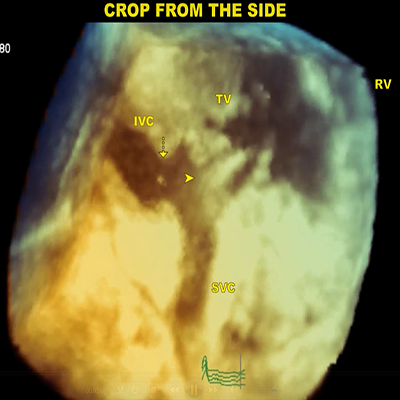
|
3D dataset cropped at an oblique angle from the top of SVC, IVC, TV, and RV
|
5 MB
|
|
|
|

|
Cropping the 3D dataset from the top initially infiltrated tissue surrounding the MV annulus
|
8 MB
|
|
|
|

|
Cropping the 3D dataset from the bottom initially the LV and pacemaker lead in the RV
|
8 MB
|
|
|
|

|
Transthoracic parasternal long axis view
|
309 KB
|
|
|
|

|
Transthoracic apical 4 chamber view
|
357 KB
|
|
|
|

|
Transthoracic apical 2 chamber view
|
402 KB
|
|
|
|

|
RV enlargement with a large well-circumscribed mass
|
210 KB
|
|
|
|

|
Echodensity
|
428 KB
|
|
|
|

|
Intraoperative surgery
|
6 MB
|
|
|
|

|
2D parasternal long-axis view
|
597 KB
|
|
|
|

|
2D apical 4-chamber view
|
432 KB
|
|
|
|

|
2D parasternal short axis view
|
317 KB
|
|
|
|

|
3D transthoracic echocardiography
|
451 KB
|
|
|
|

|
3D parasternal short axis view
|
246 KB
|
|
|
|

|
Two-dimensional echocardiography
|
372 KB
|
|
|
|

|
Two-dimensional echocardiography
|
359 KB
|
|
|
|

|
Two-dimensional echocardiography
|
1 MB
|
|
|
|

|
Doppler study
|
385 KB
|
|
|
|

|
Saline contrast echo: short axis
|
876 KB
|
|
|
|

|
Saline contrast echo: apical 4 chambers view
|
910 KB
|
|
|
|

|
3D echocardiography: LV apical view
|
1 MB
|
|
|
|

|
3D echocardiography: view from mitral plane
|
501 KB
|
|
|
|

|
3D echocardiography: view from apex
|
498 KB
|
|
|
|

|
Post-surgical echocardiogram short axis view
|
327 KB
|
|
|
|

|
Post-surgical echocardiogram apical four chambers view
|
1 MB
|
|
|
|

|
Transthoracic 2D echocardiography
|
221 KB
|
|
|
|

|
Transthoracic 2D echocardiography
|
396 KB
|
|
|
|

|
Transthoracic 2D echocardiography
|
429 KB
|
|
|
|

|
Transthoracic 2D echocardiography
|
431 KB
|
|
|
|

|
Cropped 3D-TTE
|
383 KB
|
|
|
|

|
Cropped 3D-TTE
|
1 MB
|
|
|
|

|
Zoomed view of the mass
|
333 KB
|
|
|
|

|
Cropped 3D-TTE: Zoomed view of the mass
|
528 KB
|
|
|
|

|
Intraoperative transesophageal echocardiography
|
2 MB
|
|
|
|

|
Zoomed 3D view
|
311 KB
|
|
|
|

|
Zoomed view of the mass closely packed dense echolucent areas
|
416 KB
|
|
|
|

|
Zoomed view of the mass closely packed dense echolucent areas
|
527 KB
|
|
|
|

|
2D TTE multiple mobile masses in the right and left ventricles
|
922 KB
|
|
|
|

|
2D TTE
|
2 MB
|
|
|
|

|
2D Doppler echocardiography
|
1 MB
|
|
|
|

|
Transthoracic apical 4 chamber view
|
348 KB
|
|
|
|

|
Transthoracic short axis view
|
322 KB
|
|
|
|

|
2D TTE
|
384 KB
|
|
|
|

|
Color Doppler laminar flow signals
|
1 MB
|
|
|
|
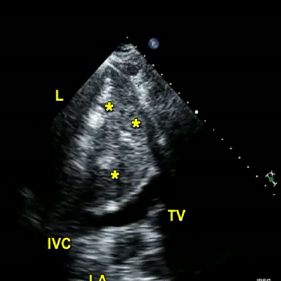
|
2D TTE
|
345 KB
|
|
|
|

|
Cystic mass in the RA
|
1 MB
|
|
|
|

|
Mass turning out to be a dilated AO
|
1 MB
|
|
|
|

|
Mild-to-moderate aortic regurgitation
|
2 MB
|
|
|
|

|
Dilated AO viewed
|
546 KB
|
|
|
|

|
Aortic regurgitation
|
318 KB
|
|
|
|

|
Subcostal view cystic structure
|
559 KB
|
|
|
|

|
Echo-free space posterior to LV
|
1 MB
|
|
|
|

|
Bubble echoes in the space (stomach, S)
|
2 MB
|
|
|
|

|
Subcostal examination in an adult patient presenting with palpitations
|
411 KB
|
|
|
|

|
Bubble studies
|
8 MB
|
|
|
|

|
Bubble studies
|
2 MB
|
|
|
|

|
Bubble studies
|
3 MB
|
|
|
|

|
2D TTE right parasternal examination
|
2 MB
|
|
|
|

|
2D TTE right parasternal examination
|
404 KB
|
|
|
|

|
2D TTE right parasternal examination
|
434 KB
|
|
|
|

|
2D TTE right parasternal examination
|
1 MB
|
|
|
|

|
2D TTE right parasternal examination
|
1 MB
|
|
|
|

|
3D TTE right parasternal examination
|
197 KB
|
|
|
|

|
Color Doppler view
|
6 MB
|
|
|
|

|
Color Doppler view
|
533 KB
|
|
|
|

|
Secundum ASD
|
256 KB
|
|
|
|

|
Parasternal long-axis RV inflow view from the transthoracic echo
|
804 KB
|
|
|
|

|
Parasternal long-axis RV inflow view from the transthoracic echo
|
641 KB
|
|
|
|

|
Intraoperative transesophageal echocardiogram
|
305 KB
|
|
|
|

|
Postoperative transthoracic echocardiogram
|
102 KB
|
|
|
|

|
Transesophageal echocardiogram
|
647 KB
|
|
|
|

|
Left parasternal view
|
515 KB
|
|
|
|

|
Subcostal view
|
531 KB
|
|
|
|

|
Right parasternal examination
|
1 MB
|
|
|
|

|
Right parasternal examination
|
5 MB
|
|
|
|

|
Right parasternal approach
|
411 KB
|
|
|
|

|
2D TTE
|
2 MB
|
|
|
|

|
2D TTE
|
614 KB
|
|
|
|

|
Transthoracic echocardiography
|
2 MB
|
|
|
|
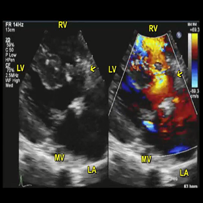
|
Parasternal long axis view 2-D and color Doppler
|
521 KB
|
|
|
|

|
Short axis color Doppler
|
482 KB
|
|
|
|

|
PLAX view
|
355 KB
|
|
|
|

|
PLAX view
|
354 KB
|
|
|
|

|
4C view
|
225 KB
|
|
|
|

|
4C view
|
357 KB
|
|
|
|

|
RVOT SAX (SAX–Aortic level)
|
346 KB
|
|
|
|

|
RVOT SAX (SAX–Aortic level)
|
166 KB
|
|
|
|

|
Post VSD Repair PLAX View
|
339 KB
|
|
|
|

|
Post VSD Repair PLAX View
|
87 KB
|
|
|
|

|
Color Doppler: apical 4-chamber view
|
937 KB
|
|
|
|

|
Transesophageal study
|
628 KB
|
|
|
|

|
Subcostal view
|
269 KB
|
|
|
|

|
Two-dimensional transthoracic echocardiography
|
158 KB
|
|
|
|

|
Two-dimensional transesophageal
|
1 MB
|
|
|
|

|
High velocity VSD flow
|
817 KB
|
|
|
|

|
Continuous high velocity flow
|
1 MB
|
|
|
|

|
LV-pulmonic plane
|
1 MB
|
|
|
|

|
LV-pulmonic plane
|
1 MB
|
|
|
|

|
Suprasternal examination
|
1 MB
|
|
|
|

|
Continuous wave Doppler
|
830 KB
|
|
|
|

|
3D TTE
|
1 MB
|
|
|
|

|
Three-dimensional transthoracic echocardiographic
|
1 MB
|
|
|
|

|
Live three-dimensional transthoracic echocardiographic
|
1 MB
|
|
|
|

|
Live three-dimensional transthoracic echocardiographic
|
2 MB
|
|
|
|

|
Transthoracic echocardiography parasternal short-axis view
|
1 MB
|
|
|
|

|
Transthoracic echocardiography parasternal short-axis view
|
1 MB
|
|
|
|

|
Transthoracic echocardiography
|
856 KB
|
|
|
|

|
Transthoracic echocardiography
|
1 MB
|
|
|
|

|
Multiplane transesophageal echocardiography
|
699 KB
|
|
|
|

|
Multiplane transesophageal echocardiography
|
1 MB
|
|
|
|

|
Right coronary angiogram
|
2 MB
|
|
|
|

|
Catheter
|
1 MB
|
|
|
|

|
Catheter
|
1 MB
|
|
|
|

|
Color Doppler flow in transthoracic subcostal view
|
1 MB
|
|
|
|

|
Transesophageal four-chamber view
|
733 KB
|
|
|
|

|
Parasternal long-axis view
|
414 KB
|
|
|
|

|
Parasternal long-axis view
|
463 KB
|
|
|
|

|
Apical 4-chamber view
|
489 KB
|
|
|
|

|
Apical 4-chamber view
|
482 KB
|
|
|
|

|
Apical 2-chamber view
|
513 KB
|
|
|
|

|
2D TTE: apical 4-chamber view
|
727 KB
|
|
|
|

|
2D TTE: apical 2-chamber view
|
723 KB
|
|
|
|

|
2D TEE: 2-chamber view
|
392 KB
|
|
|
|

|
2D TEE: 4-chamber view
|
384 KB
|
|
|
|

|
2D TEE: 4-chamber view
|
398 KB
|
|
|
|

|
3D TEE
|
935 KB
|
|
|
|

|
2D TEE: aortic short-axis view
|
1 MB
|
|
|
|

|
Transesophageal echocardiography
|
548 KB
|
|
|
|

|
Transesophageal echocardiography
|
603 KB
|
|
|
|

|
Transthoracic echocardiogram
|
265 KB
|
|
|
|

|
Transesophageal echocardiogram
|
289 KB
|
|
|
|

|
Three dimensional echocardiogram
|
5 MB
|
|
|
|

|
Three dimensional multiplanar views
|
1 MB
|
|
|
|

|
Parasternal long axis view with color Doppler
|
192 KB
|
|
|
|

|
Parasternal short axis view with color Doppler
|
420 KB
|
|
|
|

|
Subcostal view at the level of the great arteries
|
525 KB
|
|
|
|

|
Parasternal short axis view
|
656 KB
|
|
|
|

|
Subcostal short axis view
|
619 KB
|
|
|
|

|
Parasternal long axis view
|
561 KB
|
|
|
|

|
Parasternal long short axis view
|
578 KB
|
|
|
|

|
Cine aortic valve (SSFP sequence)
|
153 KB
|
|
|
|

|
Cine 4 chamber view (SSFP sequence)
|
318 KB
|
|
|
|

|
LVOT view (SSFP sequence)
|
143 KB
|
|
|
|

|
Parasternal long axis view
|
533 KB
|
|
|
|

|
Apical 4-chamber view
|
427 KB
|
|
|
|

|
Apical 4-chamber view
|
500 KB
|
|
|
|
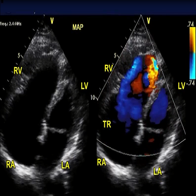
|
Apical 4-chamber view
|
519 KB
|
|
|
|

|
Illustrates LV noncompaction with a noncompaction index
|
856 KB
|
|
|
|

|
Plane velocity encoded cine imaging sequence
|
485 KB
|
|
|
|

|
LV short axis view
|
864 KB
|
|
|
|

|
Apical 4-chamber view
|
6 MB
|
|
|
|

|
Apical 4-chamber view
|
5 MB
|
|
|
|

|
Magnetic resonance imaging (MRI)
|
1 MB
|
|
|
|

|
2D echo
|
2 MB
|
|
|
|

|
2D echo
|
1 MB
|
|
|
|

|
Color Doppler echocardiography
|
1 MB
|
|
|
|

|
3D TTE 4-chamber view with focus on the tricuspid valve
|
217 KB
|
|
|
|

|
Transthoracic 4-chamber view with focus on the mitral valve
|
394 KB
|
|
|
|

|
Transthoracic apical 4-chamber view
|
220 KB
|
|
|
|

|
3D TTE 4-chamber view
|
223 KB
|
|
|
|

|
Transthoracic apical 4-chamber view with color Doppler
|
606 KB
|
|
|
|

|
3D TTE 4-chamber view
|
109 KB
|
|
|
|

|
3D TTE apical 4-chamber view with color Doppler
|
183 KB
|
|
|
|

|
Parasternal long-axis view
|
3 MB
|
|
|
|

|
Parasternal short-axis view
|
3 MB
|
|
|
|

|
Parasternal short-axis view
|
3 MB
|
|
|
|

|
Apical 4-chamber view
|
2 MB
|
|
|
|

|
Apical 4-chamber view
|
2 MB
|
|
|
|

|
Apical 4-chamber view
|
2 MB
|
|
|
|

|
SVC-IVC baffle
|
928 KB
|
|
|
|

|
SVC-IVC baffle leak
|
14 MB
|
|
|
|

|
Apical four-chamber view
|
392 KB
|
|
|
|

|
Apical five-chamber view
|
338 KB
|
|
|
|

|
Apical 4-chamber view
|
1 MB
|
|
|
|

|
3D TEE full volume acquisition
|
639 KB
|
|
|
|

|
3D TEE full volume acquisition
|
669 KB
|
|
|
|

|
3D TEE full volume acquisition
|
628 KB
|
|
|
|

|
3D TEE full volume acquisition
|
661 KB
|
|
|
|

|
3D TEE full volume acquisition
|
700 KB
|
|
|
|

|
3D TEE full volume acquisition
|
849 KB
|
|
|
|

|
3D TEE full volume acquisition
|
829 KB
|
|
|
|

|
Tricuspid valve replacement
|
1 MB
|
|
|
|

|
Transesophageal echocardiography
|
3 MB
|
|
|
|

|
Transthoracic echocardiogram, parasternal long-axis view
|
2 MB
|
|
|
|

|
Transthoracic echocardiogram, modified apical 3 chamber view
|
2 MB
|
|
|
|

|
Transesophageal echocardiogram, midesophageal short axis views
|
3 MB
|
|
|
|

|
Transesophageal echocardiogram, long-axis views
|
2 MB
|
|
|
|

|
Transesophageal echocardiogram, long-axis view
|
2 MB
|
|
|
|

|
Pulmonary regurgitation
|
307 KB
|
|
|
|

|
Parasternal short axis bidimensional and color flow views at the level of great arteries with severe pulmonary regurgitation
|
596 KB
|
|
|
|

|
The yellow arrow shows a unicuspid pulmonary valve clearly defined by 3D TTE
|
190 KB
|
|
|
|

|
A unicuspid pulmonary valve was found by 3D TTE view of the pulmonary valve (yellow arrow)
|
206 KB
|
|
|
|

|
Transesophageal echocardiography
|
903 KB
|
|
|
|

|
Coronary angiography
|
899 KB
|
|
|
|

|
Aortic short axis view
|
429 KB
|
|
|
|

|
Modified four-chamber view
|
723 KB
|
|
|
|

|
Apical four-chamber view
|
579 KB
|
|
|
|

|
Color Doppler-guided continuous wave Doppler
|
836 KB
|
|
|
|

|
2D echocardiogram: right ventricular inflow view
|
629 KB
|
|
|
|

|
2D echocardiogram: apical four-chamber view
|
610 KB
|
|
|
|

|
2D echocardiogram: parasternal short-axis view
|
633 KB
|
|
|
|

|
Coronary angiogram
|
1 MB
|
|
|
|

|
Two-dimensional transthoracic echocardiography
|
1 MB
|
|
|
|
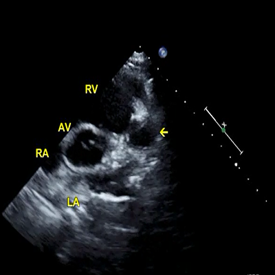
|
2D TTE
|
1 MB
|
|
|
|

|
2D TTE color Doppler
|
1 MB
|
|
|
|

|
Coronary angiogram
|
1 MB
|
|
|
|

|
Two-dimensional transthoracic echocardiography
|
794 KB
|
|
|
|

|
Two-dimensional transthoracic echocardiography
|
488 KB
|
|
|
|

|
Two-dimensional transthoracic echocardiography
|
1 MB
|
|
|
|

|
Two-dimensional transthoracic echocardiography
|
5 MB
|
|
|
|

|
Normal mitral valve nomenclature
|
714 KB
|
|
|
|

|
Mitral stenosis
|
692 KB
|
|
|
|

|
Mitral valve: atrial flutter
|
714 KB
|
|
|
|

|
Mitral valve: atrial flutter-fibrillation
|
715 KB
|
|
|
|

|
M mode examination of the left ventricle
|
524 KB
|
|
|
|

|
Left ventricle
|
149 KB
|
|
|
|

|
M mode examination of the left ventricle hypertrophy
|
226 KB
|
|
|
|

|
Stroke volume
|
648 KB
|
|
|
|

|
Discrete subaortic stenosis
|
142 KB
|
|
|
|

|
Tricuspid annular plane systolic excursion (TAPSE)
|
326 KB
|
|
|
|

|
2D TTE
|
1 MB
|
|
|
|

|
Two-dimensional transthoracic echocardiography
|
1 MB
|
|
|
|

|
Aorta short axis view
|
1 MB
|
|
|
|

|
Main pulmonary artery
|
753 KB
|
|
|
|
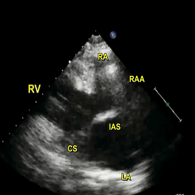
|
Right parasternal approach
|
1 MB
|
|
|
|

|
Right parasternal examination
|
496 KB
|
|
|
|

|
Crista terminalis
|
155 KB
|
|
|
|

|
Inter-atrial septum
|
174 KB
|
|
|
|

|
Right parasternal examination
|
215 KB
|
|
|
|

|
Supraclavicular approach
|
1 MB
|
|
|
|

|
Transducer is placed in the suprasternal notch and angled rightward and then left-
ward to view
|
10 MB
|
|
|
|

|
Left pulmonary artery courses inferiorly
|
2 MB
|
|
|
|

|
Right pulmonary artery (RPA) visualized
|
2 MB
|
|
|
|
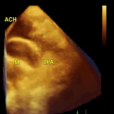
|
Three-dimensional transthoracic echocardiography
|
753 KB
|
|
|
|

|
Three-dimensional transthoracic echocardiography
|
857 KB
|
|
|
|

|
Three-dimensional transthoracic echocardiography
|
757 KB
|
|
|
|

|
Three-dimensional transthoracic echocardiography
|
787 KB
|
|
|
|

|
Three-dimensional transthoracic echocardiography
|
676 KB
|
|
|
|

|
Three-dimensional transthoracic echocardiography
|
144 KB
|
|
|
|

|
Three-dimensional transthoracic echocardiography
|
683 KB
|
|
|
|

|
Subcostal view
|
513 KB
|
|
|
|

|
2D echo
|
626 KB
|
|
|
|

|
Parasternal long-axis view of cardiac structure
|
774 KB
|
|
|
|

|
Intermittent obliteration
(A, artifact) of cardiac structures imaged in parasternal long-axis view
|
293 KB
|
|
|
|

|
Color Doppler examination
|
863 KB
|
|
|
|

|
Severe mitral valve prolapse with chordal rupture
|
522 KB
|
|
|
|
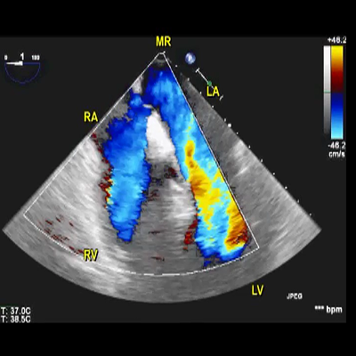
|
Severe mitral regurgitation
|
225 KB
|
|
|
|

|
MV annuloplasty systolic anterior movements
|
521 KB
|
|
|
|

|
LVOT obstruction and severe MR
|
233 KB
|
|
|
|

|
Transthoracic echocardiography
|
395 KB
|
|
|
|

|
Transthoracic echocardiography
|
408 KB
|
|
|
|

|
Transthoracic echocardiography
|
441 KB
|
|
|
|

|
Color Doppler echocardiography
|
114 KB
|
|
|
|

|
Systolic anterior motion of the mitral valve
|
594 KB
|
|
|
|

|
Aortic stenosis and significant ventricular hypertrophy
|
257 KB
|
|
|
|

|
Hypertrophic cardiomyopathy are well seen together with calcific aortic stenosis
|
498 KB
|
|
|
|

|
Pericardial and left pleural effusion
|
392 KB
|
|
|
|

|
Pericardial and left pleural effusion
|
404 KB
|
|
|
|

|
Marked swinging of the heart
|
519 KB
|
|
|
|

|
Pericardial and left pleural effusion
|
494 KB
|
|
|
|

|
Mitral valve inflow
|
319 KB
|
|
|
|

|
Right ventricle diastolic collapse
|
396 KB
|
|
|
|

|
Minimal collapse of the IVC
|
357 KB
|
|
|
|

|
Echo of cardiac tamponade prominent RV diastolic collapse
|
1 MB
|
|
|
|

|
Aortic short axis: right atrium wall collapse
|
270 KB
|
|
|
|

|
Apical 4 chamber views: right atrium wall collapse
|
405 KB
|
|
|
|

|
Fibrin band in the effusion
|
289 KB
|
|
|
|

|
Marked swinging of the heart in the short axis views toward the apex
|
288 KB
|
|
|
|

|
Subcostal view
|
375 KB
|
|
|
|

|
Dilated non-collapsing IVC
|
372 KB
|
|
|
|

|
Complete resolution of pericardial effusion with a collapsing normal-sized IVC. Ascites is noted
|
445 KB
|
|
|
|

|
Pericardial effusion with a collapsing normal-sized IVC
|
537 KB
|
|
|
|

|
Pericardial effusion with a collapsing normal-sized IVC
|
182 KB
|
|
|
|

|
2D echocardiography
|
456 KB
|
|
|
|

|
2D echocardiography
|
1 MB
|
|
|
|

|
2D echocardiography
|
589 KB
|
|
|
|

|
2D echocardiography
|
810 KB
|
|
|
|

|
Apical four chamber view: tricuspid valve fibroelastoma
|
170 KB
|
|
|
|

|
Parasternal long axis view: fibroelastoma on the aortic valve
|
1 MB
|
|
|
|

|
Aortic short axis view
|
1 MB
|
|
|
|

|
2DTTE: Arrows in the movies point to masses in RV wall
|
362 KB
|
|
|
|

|
2DTTE: Arrows in the movies point to masses in RV wall
|
351 KB
|
|
|
|

|
2DTTE: Arrows in the movies point to masses in RV wall
|
357 KB
|
|
|
|

|
2D TTE: large 4 × 2.5 cm mass (arrows) in LV with lateral wall hypokinesis and low normal systolic function were noted.
|
420 KB
|
|
|
|

|
2D TTE: mass (arrows) in LV with lateral wall hypokinesis and low normal systolic function were noted.
|
340 KB
|
|
|
|

|
2D TTE: large 4 × 2.5 cm mass (arrows) in LV with lateral wall hypokinesis and low normal systolic function were noted.
|
420 KB
|
|
|
|

|
Parasternal long-axis view: AV prolapse
|
204 KB
|
|
|
|

|
Parasternal short axis view
|
438 KB
|
|
|
|

|
Parasternal short axis view
|
430 KB
|
|
|
|

|
Moderately dilated ascending aorta
|
415 KB
|
|
|
|

|
Parasternal long axis view
|
448 KB
|
|
|
|

|
Long segment of SVC together with right (RIV) and left (LIV) innominate veins joining to form the SVC are visualized
|
422 KB
|
|
|
|

|
Right parasternal examination: Normal RCA in the right atrioventricular annulus (arrow head)
|
418 KB
|
|
|
|

|
Right parasternal window
|
171 KB
|
|
|
|
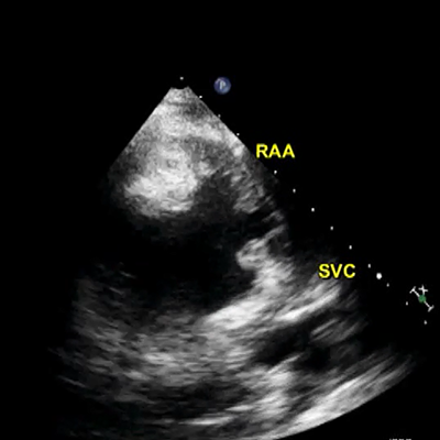
|
Right parasternal window
|
405 KB
|
|
|
|

|
Pericardial effusion
|
422 KB
|
|
|
|

|
Subcostal examinattion
|
156 KB
|
|
|
|

|
3D TTE
|
138 KB
|
|
|
|

|
Parasternal short axis view
|
186 KB
|
|
|
|

|
Parasternal short axis view
|
539 KB
|
|
|
|

|
Parasternal short
axis views
|
566 KB
|
|
|
|

|
Intraoperative 2DTEE
|
199 KB
|
|
|
|

|
Intraoperative 2DTEE
|
207 KB
|
|
|
|

|
Intraoperative 2DTEE
|
123 KB
|
|
|
|

|
Two-dimensional transthoracic echocardiography
|
138 KB
|
|
|
|

|
Transthoracic echocardiography
|
410 KB
|
|
|
|

|
Transthoracic echocardiography
|
594 KB
|
|
|
|

|
Transthoracic echocardiography
|
444 KB
|
|
|
|

|
Transthoracic echocardiography
|
378 KB
|
|
|
|

|
Transthoracic echocardiography
|
430 KB
|
|
|
|

|
Transthoracic echocardiography
|
359 KB
|
|
|
|

|
Transthoracic echocardiography
|
467 KB
|
|
|
|

|
Long axis view of LV
|
421 KB
|
|
|
|

|
Short axis view of LV
|
674 KB
|
|
|
|

|
Doppler of mitral valve
|
430 KB
|
|
|
|
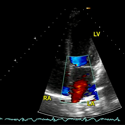
|
Doppler of mitral valve
|
648 KB
|
|
|
|

|
Utility of agited saline contrast to unmask hepatopulmonary syndrome
|
672 KB
|
|
|
|

|
2D TTE
|
3 MB
|
|
|
|
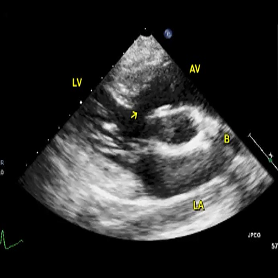
|
Transthoracic echocardiography
|
839 KB
|
|
|
|

|
Transthoracic echocardiography
|
802 KB
|
|
|
|

|
Transthoracic echocardiography
|
247 KB
|
|
|
|

|
Transthoracic echocardiography
|
831 KB
|
|
|
|

|
Transthoracic echocardiography
|
492 KB
|
|
|
|

|
Transthoracic echocardiography
|
839 KB
|
|
|
|

|
Transthoracic echocardiography
|
486 KB
|
|
|
|

|
Transthoracic echocardiography
|
836 KB
|
|
|
|

|
Dilated coronary sinus
|
739 KB
|
|
|
|

|
Transesophageal probe posteriorly the descending
thoracic aorta
|
807 KB
|
|
|
|

|
Dilated circumflex artery with turbulent flow
|
508 KB
|
|
|
|

|
TTE Parasternal long axis view
|
732 KB
|
|
|
|

|
TTE parasternal short axis view.
|
343 KB
|
|
|
|

|
TTE parasternal short axis view
|
319 KB
|
|
|
|

|
Two-dimensional transthoracic echocardiography
|
247 KB
|
|
|
|

|
Two-dimensional transesophageal echocardiography
|
431 KB
|
|
|
|

|
Two-dimensional transesophageal echocardiography
|
488 KB
|
|
|
|

|
Two-dimensional transesophageal echocardiography
|
489 KB
|
|
|
|

|
Valves were structurally normal with insignificant regurgitation
|
1 MB
|
|
|
|

|
2D TTE
|
1 MB
|
|
|
|
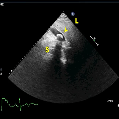
|
Stone in the gallbladder
|
720 KB
|
|
|
|

|
Stone in the gallbladder
|
341 KB
|
|
|
|

|
Echocardiogram of echodensity in the inferolateral wall
|
752 KB
|
|
|
|

|
Echocardiogram of echodensity in the inferolateral wall
|
559 KB
|
|
|
|

|
Echocardiogram of echodensity in the inferolateral wall
|
740 KB
|
|
|
|

|
Cardiac MRI
|
253 KB
|
|
|
|

|
Cardiac MRI
|
302 KB
|
|
|
|

|
Cardiac MRI
|
322 KB
|
|
|
|

|
Cardiac MRI
|
892 KB
|
|
|
|

|
Two dimensional transthoracic echo
|
3 MB
|
|
|
|

|
Two dimensional transthoracic echo
|
1 MB
|
|
|
|

|
Parasternal right ventricular inflow view
|
2 MB
|
|


